Page is loading ...

RPA II™ (Cat. # 1410)
Instruction Manual
I. Introduction . . . . . . . . . . . . . . . . . . . . . . . . . . . . . . . . . . . . . . . . . . . . . . . . . . . . . . . 1
A. Background
B. Storage and Stability
C. Reagents Provided with the RPA II™ Kit
D.Materials not Provided with the Kit
E. Related Products Available from Ambion
II. RPA II Protocol . . . . . . . . . . . . . . . . . . . . . . . . . . . . . . . . . . . . . . . . . . . . . . . . . . . . . 7
A. Before you start
B. Hybridization of Probe and Sample RNA
C. RNase Digestion of Hybridized Probe and Sample RNA
D. Separation and Detection of Protected Fragments
E. Tips for Multi-Probe RPAs and Use of Internal Controls
III. Optimization and Troubleshooting . . . . . . . . . . . . . . . . . . . . . . . . . . . . . . . . . . . . 18
A. Running the Positive Control Reaction
B. Optimization of the RPA
C. Troubleshooting Faint or Absent Protected Fragment Bands
D. Protected Fragments that are Smeared or Consist of a Ladder of Bands
E. Full Length Probe is Seen in all Lanes
F. Aberrant, Pointed or Smeared Bands
G. Radioactive Material That Fails to Migrate into the Gel Matrix
H.Reasons for Legitimate but Unexpected Bands on Autoradiographs
IV. Additional Procedures . . . . . . . . . . . . . . . . . . . . . . . . . . . . . . . . . . . . . . . . . . . . . . 34
A. Preparation and Purification of RNA Probes
B. Calculating Yield and Specific Activity of Radiolabeled Transcription Reactions
C. Calculating Yield of Non-Radiolabeled Transcription Reactions
D. Quantitation of mRNA
E. PCR Strategy to Add a Phage Promotor to Template DNA
F. Mapping mRNA
G. Recipes

Manual Version 0006
Literature Citation
We would appreciate if when you are describing a procedure utilizing this product in a Materials and
Methods Section for publication that you refer to it as the RPA II™ Kit.
Warranty and Liability
Ambion is committed to providing the highest quality reagents at competitive prices. Ambion war-
rants that the products meet or exceed the performance standards described in the product specifica-
tion sheets. If you are not completely satisfied with any product, our policy is to replace the product
or credit the full purchase price and delivery charge. No other warranties of any kind, expressed or
implied are provided by Ambion. Ambion’s liability shall not exceed the purchase price of the prod-
uct. Ambion shall have no liability for direct, indirect, consequential or incidental damages arising
from the use, results of use, or inability to use its products. This product is intended for research use
only. This product is not intended for diagnostic or drug purposes.
Copyright pending by Ambion, Inc. all rights reserved.
This product is covered by U.S. patent # 5422241
V. Appendix . . . . . . . . . . . . . . . . . . . . . . . . . . . . . . . . . . . . . . . . . . . . . . . . . . . . . . . . .50
A. References
B. RPA II™ Kit Specification Sheet
C. Guanidinium Thiocyanate Material Safety Data Sheet
D. Formamide Material Safety Data Sheet

I.A Background 1
Introduction
I. Introduction
A. Background
Procedure Overview
The Ribonuclease Protection Assay (RPA) is an extremely sensitive
procedure for the detection and quantitation of RNA species (usu-
ally mRNA) in a complex sample mixture of total or Poly(A)
selected RNA. For the RPA, a labeled (nonisotopic or radioactive)
RNA probe is synthesized that is complementary to part of the tar-
get RNA to be analyzed. This is done by placing the 3' end of the
probe sequence adjacent to one of the phage polymerase promot-
ers (T3, T7, or SP6) by cloning into a plasmid vector or by using a
PCR primer that contains the promoter sequence. (See Section
IV.E. PCR Strategy to Add a Phage Promotor to Template DNA
on
page 46.) The corresponding T3, T7, or SP6 RNA polymerase is
then used to generate an antisense RNA transcript by in vitro tran-
scription. The labeled probe and sample RNA are incubated under
conditions that favor hybridization of complementary sequences.
After hybridization, the mixture is treated with ribonuclease to
degrade unhybridized probe. Labeled probe that is hybridized to
complementary RNA from the sample will be protected from ribo-
nuclease digestion, and can be separated on a polyacrylamide gel
and visualized either by autoradiography (radioactively-labeled
probes) or by a secondary detection procedure (nonisotopi-
cally-labeled probes). When the probe is present in molar excess
over the target in the hybridization reaction, the intensity of the
protected fragment will be directly proportional to the amount of
target RNA in the sample mixture. Ribonuclease protection assays
are thus analogous to S1 nuclease protection assays, but ribonu-
clease is generally acknowledged to be easier to fine-tune and less
prone to degrade double-stranded nucleic acid than S1 nuclease
(Molecular Cloning, 1989; and Friedberg, 1990). See page 3 for a
schematic overview of the RPA procedure.
Advantages of RPAs
High Sensitivity
Compared to hybridization protocols that rely on RNA bound to
a solid support (i.e. Northern blots), low abundance mRNAs are
detected more readily and quantified more accurately by using a
solution hybridization procedure such as the RPA (Frayn, 1993,
Lee and Costlow, 1987).

RPA II™
2I.A Background
Tolerant of Partially Degraded RNA
Since the probes used in the RPA are generally significantly
shorter than the mRNA species being detected, the target RNA
preparation can be less than completely intact. Breaks in the tar-
get RNA that occur outside the region that hybridizes to the
probe will have no effect on the RPA, but would result in loss of
signal on Northern blots.
Multiple Target Analysis
Of the methods employed for RNA quantitation, RPA analysis is
the best choice for simultaneous detection of multiple targets in a
given sample. Detection of multiple targets requires only that the
probes protect RNA fragments that differ in size such that they can
be separtated on a denaturing polyacrylamide gel (Hobbs 1993).
Mapping Studies
Due to the high resolution of the acrylamide gel system used to
analyze the protected fragments, RPAs are well-suited for mapping
positions of internal and external junctions in mRNA, for example
transcription initiation and termination sites and intron
/
exon
boundaries (Kekule 1990, Melton 1984, and Calzone 1987).
Versatility
The RPA is versatile in that it can be used to discriminate between
related targets or to simultanouesly detect similar targets in a sam-
ple. To distinguish between related targets, probes are designed
to span sub-regions of relatively higher sequence divergence
(Brown et al., 1993). In this way, RNase protection can be used
to distinguish between different mRNAs coded by genes of mul-
tigene families which cross-hybridize to show a single band on
Northern blots. Conversely, to minimize the effect of probe/tar-
get sequence differences, for example when using a rat probe to
detect a heterologous mouse mRNA, the probe sequence and/or
the RNase digestion conditions can be adjusted to minimize
cleavage of mismatches.
Ambion’s RPA II™ Kit Simplifies RPAs
Ribonuclease protection assays have acquired a reputation for
being difficult to set up and optimize. The Ambion RPA II Kit is
designed to avoid many of the problems associated with ribonu-
clease protection assays, and to provide simplicity while still allow-
ing flexibility for experimental optimization. The RPA II Kit
differs from published procedures in several respects providing
greater sensitivity, as well as being faster, easier to use, and less
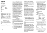
labor intensive. Figure 1 on page 3 provides an overview of the
RPA II procedure.

I.A Background 3
Introduction
Note that the proteinase K and phenol-chloroform steps of pub-
lished procedures have been eliminated and have been replaced
with a single precipitation and RNase inactivation step. This means
that the entire procedure can be performed in a single tube. This
assay is also completely compatible with nonisotopically-labeled
probes, such as those generated by Ambion's Psoralen-Biotin Kit.
The following sections include the experimental protocol as well as
information on optimization, running the positive control reaction
and troubleshooting. We encourage you to read the entire manual
if you are using the RPA II Kit for the first time.
Figure 1. RPA II™ Procedure
Denature and Hybridize
(2 – 48 hours)
Digest with RNase
(30 minutes)
Denaturing PAGE
Analysis
Combine and
Coprecipitate
Protected Fragment
Full Length Probe
Simultaneously Precipitate RNA
and Inactivate RNase
(20 minutes)
Total
RNA Antisense
Probe

RPA II™
4I.B Storage and Stability
B. Storage and Stability
The RPA II Kit should be stored at -20˚C in a non frost-free
freezer. Mouse Liver RNA can also be stored at -70˚C. A properly
stored kit is guaranteed for 6 months from the date received.
C. Reagents Provided with the RPA II™ Kit
The kit contains reagents for 120 assays.
Amount Component
2.8 ml Hybridization Buffer:
80% deionized formamide
100 mM sodium citrate pH 6.4
300 mM sodium acetate pH 6.4
1 mM EDTA
30 ml RNase Digestion Buffer
0.5 ml Yeast RNA: 5mg/ml; isolated from torulla yeast and sheared to an average size of
~300 bases
45 ml RNase Inactivation/Precipitation Solution
1.4 ml Gel Loading Buffer II: This is a 1-2X gel loading solution that can be used for
denaturing polyacrylamide gels, non-denaturing polyacrylamide gels, and TBE
agarose gels. Its composition is as follows:
95% formamide
0.025% xylene cyanol
0.025% bromophenol blue
18 mM EDTA
0.025% SDS
8 ml Probe Elution Buffer:
0.5 M ammonium acetate
1 mM EDTA
0.2% SDS
10 µl pTRI-Actin-Mouse: 5µg at 0.5 mg/ml
Linearized pTRIPLEscript DNA in TE buffer (10 mM Tris pH 8, 1 mM
EDTA); contains 250 bp of mouse ß-actin in the antisense orientation relative
to tandem SP6, T7, and T3 promoters. It yields 334 nt, 304 nt, and 276 nt
transcripts from SP6, T7, and T3 polymerases, respectively.
100 µl Mouse Liver RNA:
50 µg of mouse liver total RNA, at a concentration of 0.5 mg/ml in 0.1 mM
EDTA.

I.D Materials not Provided with the Kit 5
Introduction
D. Materials not Provided with the Kit
•
DNA template and reagents for preparing radiolabeled or
nonisotopic RNA probe (See Section V.A. for details of probe
preparation).
•
Constant temperature incubator or heat block (42˚-45˚C and
85˚-95˚C).
•
RNAse-free 1.5 ml or polypropylene microfuge tubes
•
Microcentrifuge capable of achieving at least 10,000 x g
•
Adjustable pipettors and RNAse-free tips
•
Apparatus and reagents for preparing and running denaturing
acrylamide gels (high quality urea, acrylamide and bis-acryla-
mide, Tris-borate-EDTA buffer, ammonium persulfate,
TEMED). (See Section II.D on page 14 and Section IV.2 on
page 48 for advice on gel preparation and electrophoresis.)
•
ACS grade ethanol
•
Trichloroacetic acid - molecular biology grade
•
Pasteur pipets and bulbs
•
Disposable gloves
450 µl RNase A/RNase T1 Mix
A mixture of 250 units/ml RNase A and 10,000 units/ml RNase T1 (cloned).
(RNase A is usually measured in Kunitz units. One Kunitz unit is equal to
approximately 7.5 of Ambion’s cCMP hydrolysis units.)
500 µl RNase T1 (Cloned): 5,000 units/ml
1 ml 5M Ammonium Acetate
300 µl GlycoBlue™: 15mg/ml
Amount Component

RPA II™
6I.E Related Products Available from Ambion
E. Related Products Available from Ambion
MAXIscript
™
Kits
cat. # 1310-1326
In vitro transcription kits for the synthesis of RNA probes.
MAXIscript kits are very efficient at synthesizing full length tran-
scripts even when extremely low concentrations of limiting nucle-
otide are used.
DNase I, RNase-free
cat. # 2222, 2224
DNase I is a nonspecific endonuclease that degrades double- and
single-stranded DNA and chromatin. Ambion’s RNase-free
DNase I is of the highest purity available, and is recommended to
degrade DNA in the presence of RNA when the absence of
RNase is critical.
RNA Century™ Size
Marker and RNA
Century™-Plus Size Marker
Template Sets
cat. # s 7780 & 7782
Templates for the transcription of 100-500 and 100-1000 nt
RNA molecular weight markers. Also available as pre-transcribed
biotinylated RNAs (Cat. #s 7175, 7180)
Antisense Internal Control
Templates
cat. # see catalog
These templates for in vitro transcription consist of linearized
pTRIPLEscript™ vectors that can be used to produce antisense
RNA to messages that are expressed at relatively constant levels.
They can be transcribed using any of the common RNA poly-
merases - SP6, T7, or T3. These templates can also be used in
primer extension to make single-stranded DNA. Please see
Ambion's catalog for a complete listing.
Antisense Probe Templates
cat. # see catalog
A variety of linearized plasmid templates for transcribing anti-
sense RNA probes to selected oncogenes, cell surface proteins,
cytokines, immunoglobulins, DNA binding proteins, and apop-
tosis regulators. These templates can also be used in primer
extension to make single-stranded DNA. Please see Ambion's
catalog, or call technical services as this list is always growing
Prepared RNA
cat. # see catalog
Ambion provides high quality, total and poly(A) RNA from a
variety of mouse and rat tissues and embryos, and total and
poly(A) RNA from some human tissues. These RNAs are shown
to be intact and they are precisely quantitated.
BrightStar™ BioDetect™
cat. # 1930
Low background, high sensitivity detection kit for biotinylated
RNA and DNA probes. This nonisotopic detection system is
compatible with Northern, Southern, dot blot and nuclease pro-
tection assays.
BrightStar-Plus™
Membranes
cat. # 10100-10104
Positively-charged nylon membranes recommended for use with
Ambion’s BrightStar™ nonisotopic labeling and detection prod-
ucts. These membranes are an excellent choice for Northerns and
other blot hybridizations.

II.A Before you start 7
RPA II Protocol
II. RPA II Protocol
A. Before you start
Most researchers are acutely aware of the risk of RNase contamina-
tion, and we do not want to belabor this point or cause undue
worry. We
do not
routinely find it necessary to autoclave the micro-
centrifuge tubes used in RPA reactions if they are from unopened
bags or from bags with which care was taken to avoid contaminat-
ing the tubes. We
do
recommend that gloves be worn when han-
dling any reagents or reaction vessels. If RNase contamination of
reagents or equipment is suspected to be a problem, extra precau-
tions may be necessary, for instance autoclaving tubes and tips and
aliquoting reagents to minimize chances of introducing RNase
from exogenous sources. Care should be taken not to accidentally
contaminate the kit components with ribonuclease. To avoid con-
tamination of kit components, we recommend that pipetting traffic
from stock solutions be kept to a minimum, i.e. remove the
required amount of a reagent for a set of assays to a clean tube and
withdraw the aliquots for each assay from that vessel.
Optional Coprecipitant
GlycoBlue
is included in the RPA kit to facilitate visualization
and recovery of the protected RNA pellet at the end of the exper-
iment. It is supplied as a 100X stock solution. It should be added
to the RNase Digestion Buffer at a final concentration of 1X.
•
This can be done the first time a 10 ml bottle of RNase Diges-
tion Buffer is thawed by adding 100 µl of the 100X GlycoBlue
solution.
•
Alternatively, GlycoBlue can be added to the RNase Digestion
Buffer when preparing the working dilution of RNase. Simply
add 1/100th volume of the 100X GlycoBlue stock solution in
addition to RNase to the RNase Digestion Buffer.
•
Finally, GlycoBlue can be added to RPA experiments after the
RNase digestion, before the inactivation/precipitation step. In
a typical RPA II, add 2.2 µl 100X GlycoBlue solution to each
experimental sample tube, vortex to mix, then add 300 µl
RNase Inactivation/Precipitation Solution.

RPA II™
8II.B Hybridization of Probe and Sample RNA
B. Hybridization of Probe and Sample RNA
Amount of sample RNA
and RNA probe to use
The amount of sample RNA required will depend on the abun-
dance of the mRNA being detected and on the specific activity of
the probe.
Typically, 5 - 20 µg of total RNA is used
; there is no
lower limit to how much RNA can be used in an RPA, but the
upper limit is ~50 µg. RNA preparations can consist of total RNA
or of Poly(A) selected RNA.
We recommend using gel
purified probe or probe that
has been determined to con-
sist mainly of full-length tran-
script as assessed by gel
electrophoresis; (See Section
IV.B on page 38)
For quantitative detection of mRNA, it is important that the
labeled probe be present in molar excess over the target mRNA.
Ideally, there should be a 3 to 10 fold molar excess of probe over
target mRNA. If the abundance of the target mRNA is known,
the amount of probe required to achieve 3 to 10 fold molar
excess can be calculated.
In most cases, 150-600 pg or 2-8 x 10
4
cpm of high specific activity probe per 10 µg total RNA is suffi-
cient probe to be in molar excess for B-actin or GAPDH, both
fairly abundant mRNAs.
For example, if the target mRNA is
abundant (i.e. > 0.1% of all mRNA), then 10 µg of total cellular
RNA would contain about 0.6 fmol of the target mRNA, assum-
ing that the target mRNA is 1.5 kb long and that mRNA com-
prises 3% of total cellular RNA (1 fmol = 10
-15
moles). Thus, a
four-fold molar excess of a 300 nucleotide probe with a specific
activity of 3 x 10
8
cpm/µg would require 2.4 fmol or about 7 x
10
4
cpm, corresponding to about 240 pg. If the message is less
abundant, or less sample RNA is used in the hybridization reac-
tion, then fewer cpm of probe would be needed. If the probe is
longer than 300 nucleotides, or has a specific activity greater than
3 x 10
8
cpm/µg, then more cpm of probe will be needed to
achieve 4-fold molar excess. Further guidelines for optimizing
amounts of probe and sample RNA are given in section
III. Opti-
mization and Troubleshooting
on page 18. (See also Section
IV.B.
Calculating Yield and Specific Activity of Radiolabeled Transcrip-
tion Reactions
on page 38).
Hybridization: Standard
vs. Streamlined
Procedures
There are two alternative procedures for hybridization of probe
and sample RNA. In the standard procedure, probe and sample
RNAs are co-precipitated and resuspended in hybridization
buffer. In the streamlined procedure, small volumes of the RNAs
are added directly to hybridization buffer without an initial
coprecipitation step. We expect that most experiments will be
done according to the standard procedure and we recommend
that new users follow the standard procedure. The streamlined
procedure is appropriate for processing a large number of samples
when maximum sensitivity is not critical, and when the volume of

II.B Hybridization of Probe and Sample RNA 9
RPA II Protocol
probe and sample RNA is less than 15 µl. In the streamlined pro-
cedure, the increased volume of the hybridization reaction will
have lower salt and RNA concentrations, resulting in a decreased
hybridization rate. For example, the hybridization rate in the
streamlined protocol in a total volume of 30 µl (20 µl Hybridiza-
tion Buffer plus 7 µl sample RNA plus 3 µl probe in Probe Elu-
tion Buffer) will be about 2.25-fold slower than in the standard
20 µl hybridization protocol. The streamlined procedure saves
about 30 minutes of hands-on time (and 30 minutes of precipita-
tion/centrifugation time) but it may require longer hybridization
times and it may be less sensitive. One additional note: increasing
the ratio of Probe Elution Buffer to sample RNA may actually
speed up the apparent hybridization rates due to salt effects.
Users may want to experiment with this variable.
B.I. Standard Hybridization Procedure
1. Mix sample RNA and
labeled probe
For each experimental tube, mix labeled probe with sample RNA
in a 1.5 ml microfuge tube. Use about 150 - 600 pg or
2 - 8 x 10
4
cpm per 10 µg total or 0.6 µg poly(A+) sample RNA.
A typical experiment might include 20 tubes with different
amounts or sources of sample RNA.
2. Set up 2 control tubes
for each probe
For each different probe used, include two control tubes contain-
ing the same amount of labeled probe used for the experimental
tubes in Step 1 plus Yeast RNA equivalent to the highest amount
of sample RNA. See section
III.B. Optimization of the RPA
on
page 20 for further guidelines on setting up the initial experi-
ment.
3. Co-precipitate the
probes and sample
RNAs
Adjust the concentration of NH
4
OAc to 0.5 M with the
5 M NH
4
OAc supplied with the kit.
Add 2.5 volumes of EtOH, and mix thoroughly.
4. Place tubes in -20˚C
freezer for 15 min
This is the minimum incubation time. Samples can be left indefi-
nitely at -20˚C or at -80˚C at this step.
5. Centrifuge 15 min,
max speed
Pellet the RNAs by centrifuging at maximum speed in a micro-
centrifuge (at least 10,000 rpm) for 15 minutes, preferably at
4˚C. To make it easier to see the pellets, we suggest positioning
all the tubes the same way during the spin, for example with the
hinges of the lids facing away from the center of rotation. If this is
done, the pellets will all form directly below the hinges.

RPA II™
10 II.B Hybridization of Probe and Sample RNA
6. Discard supernatant,
air dry pellets 5 min
Remove the EtOH supernatant, taking care to avoid dislodging
the pellets. It is advisable to spin tubes a second time for just
5 seconds to collect any supernatant that was clinging to the sides
of the tube, and to remove this residual liquid with a very fine
pipet tip. Let pellets dry for 5 minutes or so on the bench. Dry-
ing in a vacuum desiccator is not recommended because it may
make resuspension difficult.
7. Resuspend pellets in
20 µl Hybridization
Buffer
After adding the Hybridization Buffer to each pellet, vortex each
tube for about 5-10 seconds, then microfuge for a few seconds to
collect the liquid at the bottom of the tube.
8. Incubate 3-4 min at
90-95˚ C
This incubation denatures the RNA and aids in its solubilization.
Vortex tubes after the incubation and microfuge briefly to collect
the contents in the bottom of the tube.
9. Incubate overnight at
42˚C
Incubate tubes at 42˚C overnight to hybridize probe to its com-
plement in the sample RNA.
The
hybridization time
can be as short as 2 hours for moder-
ately abundant messages such as
β
-actin in mouse liver RNA.
Hybridization times that yield very intense protected fragment
signals may be reduced in subsequent experiments. For accurate
quantitation, however, hybridization reactions must go essentially
to completion.
The
temperature of hybridization
can also be optimized for
certain RNAs. Higher hybridization temperatures are sometimes
beneficial; some multiprobe RPA protocols suggest 56˚C as the
hybridization temperature. (If you are using Pharmingen’s tem-
plate sets, call Ambion’s Technical Service Department and
request our protocol for using these templates with MAXIscript
and RPA II.)
To minimize or eliminate condensation around the tops of the
tubes during hybridization, they should be tightly capped and
preferably incubated in a cabinet-type incubator. Alternatively,
the tubes can be incubated in a water bath or in a water-filled heat
block.
B.II. Streamlined Hybridization Procedure
If the probe and sample RNA are in a sufficiently small volume,
20 µl of Hybridization Buffer can be added directly to the sample
RNA and probe, omitting the initial ethanol co-precipitation
which is the first step of the standard procedure. We recommend
that the volume of probe in Probe Elution Buffer be at most 3 µl

II.B Hybridization of Probe and Sample RNA 11
RPA II Protocol
and that the volume of sample RNA be no greater than 12 µl, in
order to keep the salt concentration sufficiently high in the
hybridization reaction.
1. Mix sample RNA and
labeled probe
For each experimental tube, mix labeled probe with sample RNA
in a 1.5 ml microfuge tube. Use about 150-600 pg or 2-8 x 10
4
cpm per 10 µg total or 0.6 µg poly(A+) sample RNA. A typical
experiment might include 20 tubes with different amounts or
sources of sample RNA. Note that adding more probe than this
will typically not enhance signal but may increase background.
2. Set up 2 control tubes
for each probe
For each different probe used, include two control tubes contain-
ing the same amount of labeled probe used for the experimental
tubes in Step 1 plus Yeast RNA equivalent to the highest amount
of sample RNA. (See Section III.B on page 20 for further guide-
lines for setting up the initial experiment.)
3. Add 20 µl
Hybridization Buffer to
each tube
After adding the Hybridization Buffer to each tube, mix thor-
oughly by vortexing. Centrifuge tubes briefly to collect all liquid
at the bottom of the tube.
4. Incubate 3-4 min at
90-95˚ C
This incubation denatures the RNA. Vortex tubes after the incu-
bation and microfuge briefly to collect the contents in the bottom
of the tube.
5. Incubate overnight at
42˚ C
Incubate tubes at 42˚ C for 16-48 hours to hybridize probe to its
complement in the sample RNA.
The required
hybridization time
will vary depending on the
abundance of target RNA in the sample and will be longer than
with the standard procedure. Extended incubation time may be
required to compensate for the decrease in hybridization rate due
to the increased reaction volume.
The
temperature of hybridization
can also be optimized for
certain RNAs. Higher hybridization temperatures are sometimes
beneficial; some multiprobe RPA protocols suggest 56˚C as the
hybridization temperature. (If you are using Pharmingen’s tem-
plate sets, call Ambion’s Technical Service Department and
request our protocol for using these templates with MAXIscript
and RPA II.)
To
minimize or eliminate condensation
around the tops of the
tubes during hybridization, they should be tightly capped and
preferably incubated in a cabinet-type incubator. Alternatively,
the tubes may be submerged in a water bath or water-filled heat
block.

RPA II™
12 II.C RNase Digestion of Hybridized Probe and Sample RNA
C. RNase Digestion of Hybridized Probe and Sample RNA
1. Prepare a working
dilution of RNase in
RNase Digestion Buffer
Thaw a bottle of RNase Digestion Buffer, vortex well, and
remove 200 µl x the number of assay tubes to a fresh tube. Vortex
and spin the tube of RNase A/RNase T1 Mix briefly, and add the
appropriate amount of RNase A/RNase T1 Mix to the RNase
Digestion Buffer. We recommend using a 1:100 dilution; how-
ever, the optimal concentration is somewhat template dependent
and is best determined empirically. (Section III.D on page 26
contains information on optimizing RNase concentrations.) Vor-
tex and spin the RNase mixture briefly to assure even dispersion
of the components. GlycoBlue can be mixed into the RNase
Digestion Buffer at this point. It is supplied as a 100X solution, to
be used at 1X final concentration in the RNase Digestion Buffer.
2. Add 200 µl diluted
RNase solution to each
sample RNA tube, and
to one of each pair of
yeast RNA controls
After hybridization, remove the tubes from the incubator or heat
block and centrifuge briefly if any condensation is present on the
sides or top of the tubes. Add 200 µl of the diluted RNase mix-
ture prepared in the previous step to:
•each tube containing sample RNA
•and to one of the two yeast RNA control tubes that have been
prepared for each probe in the experiment. These tubes will
serve as positive controls for the function of the RNases. They
will also show if the probe is being protected in the absence of
homologous sequence (Yeast RNA is not an appropriate con-
trol if the probe is expected to hybridize with sequences found
in yeast RNA). Ideally there should be no signal at all in this
lane of the gel.
Vortex and microfuge tubes briefly.
3. Add 200 µl RNase
Digestion Buffer
without RNase to the
remaining yeast RNA
control tube(s)
This tube(s) will serve as a control for probe integrity. It will show
the gel migration of the full length probe. If there is any unex-
pected degradation of the probe, or persistent secondary struc-
ture, it will be seen in this control. This lane should show a single
band migrating at the expected probe size.
4. Incubate 30 minutes at
37˚C
During this incubation, unprotected single-stranded RNA will be
digested. In rare cases, a lower incubation temperature may be
desirable (more information in section III.D on page 26).
5. Add 300 µl RNase
Inactivation/Precipitati
on Solution
After adding the RNase Inactivation/Precipitation Solution, vor-
tex and microfuge tubes briefly. While it is not necessary to add
additional carrier during this precipitation, adding GlycoBlue™ to

II.C RNase Digestion of Hybridized Probe and Sample RNA 13
RPA II Protocol
the digestion buffer, or to each tube just before adding the RNase
Inactivation/Precipitation Solution, will make the pellets slightly
larger and much more easily visible (see page 7). Alternatively,
1-2 µl of the Yeast RNA provided in the kit can be added to each
sample just after the addition of the RNase Inactivation/Precipi-
tation Solution to increase the size and visibility of the final pel-
lets.
Precipitation of very small fragments (less than 150 bases) can be
improved by adding 100 µl of ethanol (for fragments of at least
100 bases) or 200 µl of ethanol (for fragments of at least 50
bases), in addition to 300 µl of RNase Inactivation/Precipitation
Solution.
6. Incubate at -20˚C,
15 min
Transfer tubes to -20˚C freezer for at least 15 minutes. The
experiment can be left at -20˚C overnight or longer if desired at
this step. Extended storage of radiolabeled probes will result in
radiolysis, causing an increase in background on the final autorad-
iograph.
7. Microfuge 15 min at
top speed
Remove tubes from freezer and pellet the precipitated products
of the RNase digestions for 15 minutes at maximum speed (at
least 10,000 x g), preferably at 4˚C. To make it easier to see the
pellets, we suggest positioning all the tubes the same way during
the spin, for example with the hinges of the lids facing away from
the center of rotation. If this is done, the pellets will all form at
the back of the tubes, directly below the hinges.
8. Carefully remove all
supernatant from each
tube
RNA pellets do not adhere tightly to the walls of standard
polypropylene microcentrifuge tubes, and care must be taken to
avoid losing them when removing the supernatant after centrifu-
gation. The bulk of the supernatant can be removed by gentle
aspiration or by carefully pouring the solution out of the tubes
from the side opposite the RNA pellet. To remove the last traces
of supernatant, re-centrifuge the tubes for about 5 seconds (room
temperature is OK) and withdraw the residual supernatant with a
very fine pipet tip or a drawn-out Pasteur pipette.
Do not remove the residual fluid by vacuum-drying, because the
salts present in RNase Inactivation/Precipitation Solution will
cause aberrant migration of the protected fragment during elec-
trophoresis.

RPA II™
14 II.D Separation and Detection of Protected Fragments
D. Separation and Detection of Protected Fragments
1. Prepare a denaturing
polyacrylamide gel
(Recipe on page 48). The gel size and acrylamide concentration
will be dictated by the experiment; specifically, the number and
sizes of probes, and their relation to each other. A 5% acrylamide
gel will effectively resolve fragments of about 50-1,000 nucle-
otides. We typically run 0.75 mm thick gels, 15 cm wide x 12 cm
long, with 20 wells that are about 4 mm in width. When more
than 3 or 4 probes are used together, it may be better to use short
sequencing-type gels. (Section II.E. Tips for Multi-Probe RPAs
and Use of Internal Controls on page 15 contains more informa-
tion on multiple probes.) It is helpful to have size markers on the
gel, single-stranded RNA markers are the most accurate, but dou-
ble-stranded DNA markers can be used if it is not critical to know
the exact size of the products.
2. Resuspend pellets in
Gel Loading Buffer II
The volume of gel loading buffer used is not critical, but the best
resolution is obtained when the gel loading buffer forms a
2-3mm layer in the well; this is usually 4-10µl. Vortex vigorously
and microfuge briefly.
3. Incubate 3 min at
90-95˚C
This incubation helps to completely solubilize the RNA and
denatures it. After the incubation, vortex and microfuge again
briefly. Store the tubes on ice before loading them on the gel.
4. Load the samples and
run the gel
Rinse the urea out of the wells of the gel, and immediately load
each sample. Load only 10 to 20% of the no target/no RNase
controls compared to the experimental samples to avoid obscur-
ing the signal from adjacent lanes (the no target/no RNase con-
trols are expected to contain many more counts than the
experimental samples). Gels of the size described at the beginning
of this section should be run at about 250 volts constant voltage
or 25-30 mamps constant current. The run time is typically about
one hour Although it will vary depending on the size of the pro-
tected fragment. We typically run RPA gels until the leading dye
band (bromophenol blue) is near the bottom of the gel.
4a. Detection of
radiolabeled probes
Transfer the gel to filter paper, mark the origins and orientation
of lanes, cover with plastic wrap, and expose to X-ray film for an
appropriate length of time. We usually expose from overnight to
several days using single-side coated X-ray film (e.g. Kodak XRP)
without an intensifying screen, or for several hours to overnight
with an intensifying screen. Expose at -80˚ or -20˚C. The gel can
be re-exposed several times if necessary after allowing it to warm
up to room temperature and wiping off condensation moisture.

II.E Tips for Multi-Probe RPAs and Use of Internal Controls 15
RPA II Protocol
The gel should be stored at -80˚ or -20˚C if not re-exposed
immediately. Users may prefer to dry gels onto chromatography
paper; we do not find it necessary to dry standard 0.75 mm thick
gels. If probes were labeled with 35S instead of 32P, gels should be
dried and exposed directly to X-ray film (without plastic wrap).
Note: Gels with 35S-labeled probes will require fluorography for
maximum sensitivity, and will be about 10-fold less sensitive than
gels with 32P-labeled probes if exposed directly without using flu-
orographic techniques. Intensifying screens will not increase the
sensitivity of 35S-labeled probes.
4b. Detection of
nonisotopic probes
Transfer the gel to a positively-charged nylon membrane by elec-
troblotting, crosslink nucleic acids to the membrane, and follow
the protocol of an appropriate detection procedure for visualiza-
tion of the nonisotopic probe. (e.g. Ambion’s BrightStar BioDe-
tect Kit.)
E. Tips for Multi-Probe RPAs and Use of Internal Controls
1. Templates Quantitation of mRNA by nuclease protection assay requires that
signal resulting from undigested full-length probe not interfere
with signal from protected fragments. Additionally each of the
full-length fragments should be spaced sufficiently so that their
signals will not obscure those from other protected fragments.
When only one or two probes are being used, this is typically not
a problem; but when a large number of probes are used simulta-
neously, probe design becomes very important. If possible,
probes should be designed in such a manner that the size of the
expected protected fragments will be at least 10% different from
any other full-length probe or protected fragment.
Additionally, there is the potential problem of probe interactions.
There has been one report of a probe for 18S RNA, which was
present at relatively high levels in the hybridization, forming an
RNase resistant complex with one of the other probes in a
multi-probe RPA. This occurred via hybridization of a 20 bp
complementary region that was transcribed from the probe tem-
plates’ multiple cloning sites (Ginter et al. 1994).
2. Transcription reactions
in small volumes
Probe synthesis tends to be one of the most costly aspects of
multi-probe analysis. Probe synthesis costs can be reduced by
performing transcription reactions in smaller volumes. A 2-5 µl
transcription will usually provide enough gel purified probe to
perform dozens of nuclease protection assays. Several small tran-

RPA II™
16 II.E Tips for Multi-Probe RPAs and Use of Internal Controls
scription reactions can be conveniently assembled by generating a
master mix comprising transcription buffer, NTPs, and poly-
merase and aliquoting appropriate volumes of master mix into
individual tubes containing the various templates to be tran-
scribed.
3. Reduce radiolytic
probe decay
Radiolabeled transcripts degrade relatively quickly due to radi-
olytic decay. High background and even aberrant bands can
result from using radiolabeled probes that are several days old.
For the best results, probes should be gel-purified, eluted, and
assembled in hybridization reactions on the same day.
4. Equalize protected
fragment intensities
a. Radiolabeled Probes
When targets that vary widely in abundance are assayed simul-
taneously, it is important to equalize the intensities of the pro-
tected fragment bands by adjusting the specific activities of the
probes. This is done by adding unlabeled NTP to dilute the
corresponding labeled NTP in the transcription reaction, thus
lowering probe specific activity. For instance, when transcrib-
ing probes for two messages that differ 200-fold in abundance,
unlabeled UTP should be added to the transcription reaction
for the more abundant mRNA at a final molar concentration
that is 200-fold greater than the labeled UTP. The table
below provides a starting point for reducing specific activities
of probes to common internal controls, further optimization
of probe specific activity may be required to achieve ideal
results. For each probe, add the same number of cpm to the
hybridization reactions so that all of the full-length probes will
be visible in the no target/no RNase control lane.
b. Nonisotopically labeled probes
When non-isotopic labeling is used, the specific activities of
the probes should be adjusted by titrating non-isotopically
labeled with unlabeled probe at ratios of 1:160,000 for riboso-
mal RNAs, 1:200 for GAPDH and ß-actin, and 1:20 for cyclo-
philin.
Internal Controls
labeled:unlabeled
nucleotide [α-32P]UTP UTP specific activity
Ribosomal RNAs (18S and 28S) 1:160,000 0.01 µl 2 µl 2.5 x 10⁴ cpm/µg
Moderate Abundance Targets
(ß-actin and GAPDH) 1:200 4 µl 1 µl 2 x 10⁷ cpm/µg
Low Abundance Targets (cyclo-
philin) 1:20 4 µl 0.1 µl 2 x 10⁸ cpm/µg

II.E Tips for Multi-Probe RPAs and Use of Internal Controls 17
RPA II Protocol
5. Decrease probe
interactions by raising
the hybridization
temperature
Occasionally, cross-hybridization between probes (observed as a
distinct band in the no target/+ RNase control lane as well as in
the sample RNA lanes) or between a probe and non-target RNA
can affect detection or quantitation. Often, increasing hybridiza-
tion stringency by raising the hybridization temperature to 56˚C
or 68˚C can eliminate the cross-hybridization without affecting
probe:target interactions.

RPA II™
18 III.A Running the Positive Control Reaction
III. Optimization and
Troubleshooting
A. Running the Positive Control Reaction
The positive control reaction included with the RPA II Kit consists
of Mouse Liver sample RNA and pTRI-Actin-Mouse: a DNA tem-
plate for transcription of an antisense mouse β-actin RNA probe.
1. Probe Preparation
To synthesize a radiolabeled positive control probe, use 1 µl of
pTRI-Actin-Mouse DNA template in a 10 µl or 20 µl in vitro tran-
scription reaction containing at least 3 µm [α-
32
P]UTP or
[α-
32
P]CTP (50 µCi of 800 Ci/mmol 10 µCi/ µl) and up to 5 µM
unlabeled nucleotide corresponding to the
32
P-NTP used. (The
lower the concentration of unlabeled nucleotide, the higher the
specific activity of the transcript will be, and thus the greater the
sensitivity of the assay. For maximum sensitivity, do not add any
unlabeled form of the limiting nucleotide.) Any of the three com-
mon phage polymerases can be used in the transcription reaction
because pTRI-Actin-Mouse has tandem SP6, T7, and T3 promot-
ers. The sizes of the transcripts are 334 bases, 304 bases, and
276 bases when pTRI-Actin-Mouse is transcribed with SP6, T7,
and T3 polymerases, respectively. Each of these probes will protect
245 bases of the mouse β-actin mRNA. It is recommended that the
probe be treated with DNase to remove template, and that it be gel
purified for use in the control RPA (section IV.A.5 on page 36).
2. RPA Setup
Use about 4 x 10
4
cpm of β-actin probe with several different
amounts of mouse liver RNA, for example 2.5 µl, 5 µl, 10 µl, and
20 µl (corresponding to 1.25 µg, 2.5 µg, 5 µg, and 10 µg). Include
two yeast RNA control hybridizations with the same amount of
probe and 2 µl Yeast RNA. Follow the protocol for doing the RPA
outlined in the previous sections; hybridize overnight at 42˚C, and
use RNase A/RNase T1 Mix at a 1:100 dilution.
3. Expected Results
Protected fragments of 245 bases should be seen in the reactions
containing Mouse Liver RNA, and the protected fragments should
be more intense with increasing amounts of Mouse Liver RNA.
The no target/+RNase lane should have no signal, and the no tar-
get/no RNase lane should show a band corresponding to full
length probe.
/