National 131 User manual

Type
User manual

This manual is also suitable for

National 131 is a versatile compound microscope with brightfield illumination, suitable for educational and basic laboratory applications in biology, chemistry, and other fields. It features a 360-degree rotatable monocular head, allowing multiple users to observe specimens or share the microscope easily. The coarse and fine focus knobs provide precise control for sharp image focusing, while the built-in LED illuminator ensures consistent and reliable illumination of specimens.

National 131 is a versatile compound microscope with brightfield illumination, suitable for educational and basic laboratory applications in biology, chemistry, and other fields. It features a 360-degree rotatable monocular head, allowing multiple users to observe specimens or share the microscope easily. The coarse and fine focus knobs provide precise control for sharp image focusing, while the built-in LED illuminator ensures consistent and reliable illumination of specimens.

National Optical & Scientific Instruments Inc.
6508 Tri-County Parkway
Schertz, TX 78154
Phone (210) 590-9010 Fax (210) 590-1104
INSTRUCTIONS FOR
MODELS 131, 132, 131-CLED & 132-CLED
COMPOUND MICROSCOPES
NOTE: These models share common features, nomenclature and
method of operation. The only difference are…
Models 131 & 132 feature electric illumination from a
tungsten bulb.
Models 131-CLED & 132-CLED feature electric illumination from a
LED bulb.
Models 132 & 132-CLED have both 30° inclined and vertical
eyepiece tubes, each supplied with WF10x
eyepiece.
Copyright © 1/2/01
National Optical & Scientific Instrument Inc.
3
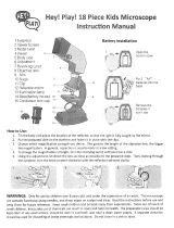
For optimum viewing satisfaction, follow these simple procedures. Nomenclature used to describe components and
controls can be identified by referring to the diagram at left.
UNPACKING
1. For Models 131 & 132, carefully remove microscope, dustcover and for Models 131-CLED & 132-CLED also remove
a “L” hex wrench. Always handle and move microscope by securely holding the arm of microscope. Avoid touching
any of the lens surfaces while handling the microscope. Dust, dirt, or fingerprints can damage the delicate lens
surfaces or adversely affect image quality.
2. Examine packing material before you discard it. Retain the styrofoam container in case you need to transport,
store, or return the microscope for service. If it becomes necessary to ship the microscope for any reason, pack it
in the styrofoam container, and then pack the styrofoam in another corrugated shipping container for optimum
protection. Use of the styrofoam alone will not provide adequate protection in transit, and will void your warranty.
DESCRIPTION OF COMPONENTS
1. EYEPIECE (ocular lens) Lens closest to the eye, magnifies the primary image formed by the objective lens. Dual
head models 132 and 132-CLED feature both an inclined eyepiece (for primary viewing) and a vertical eyepiece for
second viewer or for mounting a camera. The inclined eyepiece is equipped with a “pointer” that rotates as the
eyepiece is turned.
2. DIOPTER ADJUSTMENT (on models 132 and 132-CLED only) Permits focusing adjustment of image for any
difference in vision between primary and secondary viewers.
3. OBJECTIVE TURRET (nosepiece) Revolving turret which holds objective lenses, permits changes of magnification
by rotating different powered objective lenses into optical path.
4. OBJECTIVE LENS Lens closest to the object being viewed, forms first magnified image of the specimen.
5. STAGE CLIPS Two locked-on clips hold specimen slide in place on stage. Note: Your microscope is already drilled
and tapped to accept an optional mechanical stage. Mechanical stage replaces stage clips and permits precise,
mechanical manipulation of the specimen slide.
6. STAGE Platform of the microscope where the specimen slide is placed.
7. CONDENSER LENS An 0.65 N.A. condenser lens, fixed in center of stage, condenses light rays from substage
illumination and fills the back lens element of objective lens to improve image resolution.
8. DISC DIAPHRAGM Rotating disc located below stage, with holes of various apertures, designed to help achieve
optimum resolution of the objective lens. Larger apertures used for higher magnifications, and smaller apertures used
for lower magnifications.
9. SAFETY RACK STOP When properly adjusted, controls maximum upward travel of stage. Prevents higher power
objectives from breaking specimen slides, prevents damage to objective lenses. This stop has been pre-adjusted at
the factory.
10. FOCUSING KNOBS Coarse focusing knobs (larger knobs) located on each side of arm, raise or lower stage to
bring specimen image into focus. Fine focus knobs (smaller knobs located just below coarse focusing knobs) permit
more precise image adjustment.
11. ILLUMINATION Models 131 & 132 have a built-in substage electric illuminator and Models 131-CLED & 132-CLED
have a built-in substage electrical LED illuminator that provides constant, reliable, pre-focusing illumination.
OPERATION
1. Place microscope directly in front of you in a manner which permits you to comfortably look into the eyepiece. Note
that the head of microscope rotates 360º, permitting you to operate the microscope from the front or from the back,
whichever is most convenient for you. It also permits convenient sharing of microscope by more than one user, by
simply rotating head, without needing to move entire microscope. Most users will position the microscope with the
arm facing them so that focusing knobs are most convenient to reach.
4
2. Insert microscope plug into matched voltage outlet. On Models 131, 131-CLED & 132, flip on/off power switch located
on base to “ON” position. On Model 132-CLED, rotate on/off power switch located on base to “ON” position.
3. Rotate coarse focus knobs to move stage down (away) from objective lens as far as possible.
4. Place specimen slide, cover slip facing up, on stage with specimen centered over condenser lens in middle of stage.
5. Rotate disc diaphragm until largest aperture is positioned beneath condenser in center of stage.
6. Turn the objective turret until the 4x (smallest) objective lens “clicks” into position in the optical path. Note that each
time you change from one objective lens to another you should turn the turret until you hear the “click”, which
indicates that the lens is properly indexed in the optical path.
7. While looking through the eyepiece, rotate coarse focusing knobs until specimen comes into focus. If image does not
appear in field of view, move specimen slide slightly until image appears in field of view.
8. Adjust fine focus controls until specimen is in sharp focus.
9. Turn the disc diaphragm, observing that different apertures affect the sharpness of the image. Turn diaphragm until
sharpest possible image is obtained. Note that when turning the disc diaphragm, you should hear a “click” as each
aperture comes into proper position under the condenser lens. If aperture is not properly positioned, you will observe
shadows in the field of view when looking through the microscope.
10. Changing magnification
a. Magnification is changed by rotating objective turret until different objective lens is moved into optical path.
Always turn turret until you hear the “click”, indicating that lens is properly indexed. Otherwise, you will not be
able to see anything when looking through the microscope.
b. Standard lenses provided with your microscope are a widefield 10x eyepiece (two eyepieces supplied with
models 132 and 132-CLED). 4x. 10x, and 40x objectives. The 40x objective lens has a special spring retractable
mechanism which retracts slightly if front of lens comes in contact with the specimen slide. See chart below for
specifications on objectives.
Objective Specification Chart
Objective
N.A.
Color Code
Ring
Field of
View
Working
Distance
Magnification with
WF10X eyepiece
Din 4X
0.10
Red
4.5mm
18.5mm
40X
Din 10X
0.25
Yellow
1.8mm
6.4mm
100X
Din 40X retractable
0.65
Blue
0.45mm
0.5mm
400X
c. Also note that each objective has a color ring, which permits you to instruct changes in magnification by referring
to an easily observed color rather than to a number.
d. The microscope has been parfocalled at the factory, which allows easy change from one magnification to another,
requiring little or no adjustment of the fine focus knobs.
e. As magnification is increased, the field of view (area of specimen seen through the microscope) will decrease.
That is why it is easier to find the specific area of interest on the specimen by starting with the lowest 4x objective
lens, before increasing magnification with the 10x or 40x objective lens.
f. NOTE: Care must be taken when rotating the 40x objective into place. This lens has a spring retractable
mechanism which retracts slightly into its housing if the front of the lens strikes the specimen slide. With fine
focus adjustment at mid-range, the rack stop has been adjusted at the factory to assure the 40x lens will clear the
thickness of a normal specimen slide and cover slip. However, if the rack stop has been improperly adjusted, or if
using a thicker than normal slide or cover slip, moving the 40x lens too quickly or carelessly could cause damage
to the front lens element or to the slide.
5
g. Do not let the front lens element come into contact with a wet slide surface, as prolonged contact with any
moisture could damage the lens. If lens is exposed to moisture, promptly wipe with soft tissue to remove
moisture.
11. Diopter adjustment for models 132 and 132-CLED dual head.
These dual head models have a 30° inclined eyepiece to provide comfortable viewing for the primary viewer. They
also have a vertical eyepiece so that a second viewer can observe the image simultaneously. The primary viewer will
have focused the microscope using the coarse and fine focus knobs. To compensate for differences in vision
between primary and second viewer, second viewer may have to adjust image in vertical eyepiece by turning the
knurled diopter adjustment.
MAINTENANCE
WARNING: For your own safety, turn switch off and remove plug from power source before maintaining your
microscope. If the power cord is worn, cut or damaged in any way, have it replaced immediately to avoid
shock or fire hazard.
1. OPTICAL MAINTENANCE
Do not attempt to disassemble any lens components. Consult a microscope service technician when any repairs not
covered by instructions are needed.
Prior to cleaning any lens surface, brush dust or dirt off lens surfaces using a camel hair brush. Or use air to blow
dust and lint off surfaces. Use of compressed air in a can, available at any computer supply store, is a good source
of clean air.
Do not remove eyepieces or objective lenses to clean. Clean only the outer lens surface. Breath on lens to dampen
surface, then wipe with lens paper or tissue or use a cotton swab moistened with distilled water. Wipe lenses with a
circular motion, applying as little pressure as possible. Avoid wiping dry lens surface as lenses are scratched easily.
If excessive dirt or grease gets on lens surfaces, a small amount of Windex or distilled water can be used on a cotton
swab or lens tissue. To clean objective lenses, do not remove objectives from microscope. Clean front lens element
only, following same procedure.
NOTE: Fingerprints or other matter on the front lens element of the objective lens is the single most common reason
that you will have difficulty in focusing the microscope. Before having costly servicing done, or before returning to
National for “warranty repair”, make certain to examine the front lens element with a magnifying glass or eye loupe for
the presence of such contaminants. If a microscope is returned to National for warranty repair, and it is determined
that such contaminants are the problem, this is not covered under warranty and National will submit a cost estimate
for cleaning.
2. MECHANICAL MAINTENANCE
a. The rack stop screw has been pre-adjusted at the factory and should not require re-adjustment. However, if you
do attempt re-adjustment, note the following procedure.
Loosen round knurled locking nut by turning counter-clockwise. Then loosen stop screw. With fine focus
adjustment at mid-range, focus on standard slide until sharp image is obtained. Rotate rack stop screw in
clockwise direction until tight. Lock into position with the locking nut. A needle nose pliers might be needed to
loosen the round locking nut.
b. Coarse focus tension adjustment prevents the stage from drifting down from its own weight and causing the
image to move out of focus. This has been adjusted at the factory, but over the course of time it may loosen and
cause the stage of the microscope to slip downward on the focusing block.
The tension adjustment collar is located between arm and coarse focus knob on left side of microscope. With a
small jewelers screwdriver (models 131/132-CLED use supplied 0.90 “L” hex wrench), loosen the set screw
located in only one of the four holes on tension adjustment collar. Turn collar clockwise to tighten tension,
counter-clockwise to loosen tension. Use of a wide rubberband will provide a better grip on the tension adjustment
collar. After adjusting, tighten the set screw to lock collar in place.
6
NOTE: It is recommended that you leave the tension as loose as possible for ease of focusing, yet not so loose
that it permits the stage of microscope to drift downward from its own weight and cause the microscope to “drift
out of focus.
c. Metal parts: Use a clean, damp cloth to remove dust or dirt from metal parts, followed by a dry cloth.
3. ELECTRICAL MAINTENANCE
The extent of electrical maintenance, by other than a qualified technician, should be bulb replacement. BE CERTAIN
TO TURN SWITCHES OFF AND REMOVE PLUG FROM POWER SOURCE OUTLET BEFORE CHANGING
BULBS.
a. Changing bulb on Models 131 & 132
Make sure that illuminator field lens housing and lamp are cool before attempting to remove. Remove illuminator
field lens housing by rotating in a counter-clockwise direction. This housing is secured tightly, to prevent easy
removal by students, so a very firm grip and some strength will be required.
Note that lamp does not screw out. Remove lamp by depressing lamp slightly and rotate in a counter-clockwise
direction until it pops up slightly. Replace with a 110v 20 watt lamp with double contact, medium bayonet base.
This is a standard, commonly used replacement bulb. National #800-102. Carefully wipe lamp to insure it is
clean and free of all fingerprints. Replace field lens housing assembly.
b. Changing LED bulb on Models 131-CLED & 132-CLED
To open the illuminators field lens housing, use 0.9mm “L” hex key wrench supplied with your microscope.
Loosen hex screws on lens housing. Remove lens housing to expose LED bulb. Remove bulb by grasping the
plastic base of bulb and gently pulling straight up. Insert new LED bulb, replace lens housing and tighten hex
screws to secure lens housing in place.
c. Replacement of fuse on Models 131-CLED & 132-CLED
The fuse is located at the rear right side of microscope base. To remove fuse from holder, insert a 6mm
screwdriver blade into slot located in rear of fuse holder cap. Slightly depress and rotate screwdriver ¼ turn in
direction of arrow, release pressure on screwdriver to release fuse. Pull cap and fuse out of fuse holder. Insert
proper fuse into fuse cap. Insert fuse cap into fuse holder. Using screwdriver, rotate fuse cap assembly in
opposite direction of arrow until slot engages, depress fuse cap and rotate ¼ turn to lock into fuse holder.
7
TROUBLESHOOTING
PROBLEM
SOLUTION
Light fails to operate.
Have qualified service technician
repair outlet.
Plug into outlet.
Replace lamp.
(131/132-CLED only)
Replace LED.
(131/132-CLED only)
Replace fuse.
Image does not remain in focus
Adjust tension control.
Image will not focus
Adjust rack stop.
Place slide on stage with cover
slip up.
Use 0.17mm thick cover slip
(No.1 cover slip)
Poor resolution
(image not sharp)
Objective lenses dirty. Clean objective lenses.
Clean eyepiece lenses.
Adjust disc diaphragm.
Spots in field of view.
Eyepiece or condenser lens dirty.
Clean lens. ***
Specimen slide dirty.
Clean slide.
***Spots in field of view can also result from dirt on inside of eyepiece. It is recommended that you have
service technician clean inside of lens.
OPTIONAL ACCESSORIES AND PARTS:
#610-045R WF10x eyepiece w/reticle, 10mm/100div.
#615-045 WF15x eyepiece w/pointer
#760-045 DIN 60xR objective lens, N.A. 0.90
#800-001 Replacement LED bulb
(131-CLED & 132-CLED only)
#800-102 Replacement bulb, 115v 20 watt tungsten D.C. medium base (131 & 132 only)
#801-160 Replacement fuse, 0.5 amp, time delay
(131-CLED & 132-CLED only)
#910 Graduated mechanical stage, side mount adjustments (installed by end user)
#951 Dustcover, 16” tall x 13”, heavy vinyl with stitched seams.
#975-001 Carrying case, anodized aluminum, fabric lining, accessory pockets, Velcro straps, keyed lock. Fits all
models in series, except dual head models.
#975-002 Carrying case, anodized aluminum, fabric lining, accessory pockets, Velcro straps, keyed lock. Fits dual
head models.
#975-138 Carrying case, heavy plastic w/foam liner. Fits all models in series, except dual head models.
LIMITED LIFETIME WARRANTY
Please see our website,
www.nationaloptical.com, for complete warranty details and exclusions.
(Revised 6/24/14)
  • Page 1 1
  • Page 2 2
  • Page 3 3
  • Page 4 4
  • Page 5 5
  • Page 6 6

National 131 User manual

Type
User manual
This manual is also suitable for

National 131 is a versatile compound microscope with brightfield illumination, suitable for educational and basic laboratory applications in biology, chemistry, and other fields. It features a 360-degree rotatable monocular head, allowing multiple users to observe specimens or share the microscope easily. The coarse and fine focus knobs provide precise control for sharp image focusing, while the built-in LED illuminator ensures consistent and reliable illumination of specimens.

Ask a question and I''ll find the answer in the document

Finding information in a document is now easier with AI