Promega Corporaon · 2800 Woods Hollow Road · Madison, WI 53711-5399 USA · Toll Free in USA 800-356-9526 · 608-274-4330 · Fax 608-277-2516 7
www.promega.com TM601 · Revised 6/23
3.C. Assay Protocol (continued)
1. For each sample, standard or background control, prepare wells for measurement with and without esterase as
needed.
For Medium, Serum and Homogenized Tissue Samples:
i. Dilute samples in Cholesterol Lysis Solution to bring their cholesterol concentrations below 80μM.
Transfer 25μl of sample, standard or background control to a 96-well plate. Refer to Table 2 for dilution
recommendations.
ii. Add 25μl of Cholesterol Lysis Solution, shake briey and incubate for 30 minutes at 37°C.
For Adherent Cells and 3D Cultures:
i. Remove medium from cells in a 96-well plate. Wash cells twice with 100μl of PBS.
ii. Add 50μl of Cholesterol Lysis Solution, shake briey and incubate for 30 minutes at 37°C.
iii. If needed, dilute samples in Cholesterol Lysis Solution to bring their cholesterol concentrations below
80μM. Transfer 50μl of any diluted samples, standards or background controls to empty wells in a
96-well white-walled assay plate.
2. Add 50μl of cholesterol detection reagent with or without Esterase as prepared in Section 3.B to all wells.
3. Shake the plate for 30–60 seconds by hand or at a low rpm on a plate shaker.
4. Incubate at room temperature for 1 hour.
5. Record luminescence using a plate-reading luminometer such as the GloMax® Discover.
Note: The light signal continues to increase until all cholesterol is consumed and the signal plateaus. At any
time point the signal is directly proportional to cholesterol concentration.
6. Calculate free and total cholesterol by comparison of the luminescence of samples and standards assayed
under the same conditions (See Figure 3).
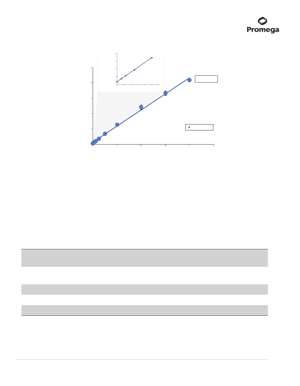
Analyte (µM)
Luminescence (RLU)
0206040 80 100
6,000,000
4,000,000
8,000,000
10,000,000
2,000,000
0
16249MA
R²=0.9957
cholesterol
1,600,000
1,200,000
800,000
400,000
0
0246 12108
Figure 3. Cholesterol standard curves. Dilutions of the provided Cholesterol Standard (20mM, in ethanol) were
prepared in Cholesterol Lysis Solution, beginning with a 2μl aliquot of Cholesterol Standard into 498μl of Cholesterol
Lysis Solution. (Refer to Table 1 for concentrations of Cholesterol Standard used in standard curve.) A 50μl aliquot of
cholesterol detection reagent was added to 50μl of each standard in triplicate and the luminescence was read after
1 hour. Concentration was plotted against average RLU at each standard point and a linear curve was t. The
Cholesterol/Cholesterol Ester-Glo™ Assay can detect less than 1µM cholesterol and has an upper limit of 80µM
cholesterol. See Sections 4.A and 4.B for other ways to convert luminescence into concentration.
Table 1. Cholesterol Titration Data.
Cholesterol
(µM) 80 60 40 20 10 5 2 1 0
RLU
(thousands)
8,343 6,685 4,825 2,537 1,389 745 444 293 134
STDEV
(thousands)
229 277 285 31 35 8 72 3
CV 2.7% 4.1% 5.9% 1.2% 2.5% 1.0% 1.6% 0.7% 2.5%
S/B 62.5 50.1 36.1 19.0 10.4 5.6 3.3 2.2 1.0
S/N 2,416.5 1,928.5 1,381.2 707.4 369.6 180.1 91.5 46.8 –
Note: Coefcient of variation (CV) is 100 × STDEV/RLU. Signal-to-background ratio (S/B) is mean signal from
samples divided by the mean signal from negative controls. Signal-to-noise ratio (S/N) is the net signal (mean signal
minus mean negative control) divided by the standard deviation of the negative control.


