Page is loading ...


ADVICE
FOR
SUPERVISING
ADULTS
a)
Read
and follow the instructions, the safety rules and the
first aid information.
Keep
them for reference.
b)
The incorrect use
of
chemicals can cause
injury
and
damage
to
health. Only carry
out
those preparations
which are listed in the instructions.
c)
This Microscope
Set
is
for
use
only by children over
10
years old.
d)
Because
children's abilities vary
so
much, even within age
groups, supervising adults should exercise discretion
as
to
which preparations
are
suitable and
safe
for them.
The
instructions should enable supervisors
to
assess
any
preparation
to
establish its suitability for a particular
child.
e)
The supervising adult should discuss the warnings and
safety information
with
the children before commencing
the preparations. Particular attention should be paid
to
the
safe
handling
of
the materials in the bottles
(i.e
. the
dyeing solution), also the functionally sharp
point
on the
needle and the functionally sharp edges on the scalpel
and slicer.
f) The
area
surrounding the experiment should be kept
clear
of
any obstructions and away from the storage
of
food.
It
should be well lit, ventilated and close
to
a water
supply. A solid table with a heat-resistant
top
should be
provided.
g)
A separate tin or bucket should be used for the disposal
of
solid waste materials. Any
was
ted solution should be
poured down a drain
but
never into a sink.
h)
To
be
used solely under the strict supervision
of
adults
that
have studied the precautions given in the
experimental
set.
EXPLANATION
OF
BOTTLES
EOSIN
Powder:
Examples for
use:
WARNING:
METHYLENE BLUE
Powder:
Examples for
use:
WARNING:
To
be mixed
with
water.
For
observation
of
the stems and roots
of
plants, also observation
of
blood and
muscle cells.
Do
not
swallow.
In
case
of
accident.
Call
a Poison center or doctor immediately.
Keep
away from young children.
To
be mixed
with
water.
For
observation of:
Cellular tissue
of
plant's leaf, blade, and
stem; mushrooms, molds,and bacteria;
blood and muscl cells.
Harmful Do
not
swallow.
In
case
of
accident.
Call
a Poison center or
doctor
immediately. Keep away from young
children.
SODIUM
CHLORIDE (SALT)
Please
refer
to
last page for usage.
SHRIMPS
EGGS:
Please
refer
to
later detailed
explanation.
IMPORTANT
TELEPHONE
NUMBERS
To
be completed by
an
adult before using the kit.
Poison Center:
Hospital:
Fire Department:
Doctor:
SAFETY
INFORMATION
General First Aid Information
a)
In
case
of
eye contact:
Wash
the
eye
with
plenty
of
water,
holding the eye open
if
necessary.
Seek
immediate
medical advice.
b)
If
swallowed:
Wash
the
mouth
with
water, drink some
fresh water. Do
not
induce vomiting.
Seek
immediate
medical advice.
c)
In
case
of
inhalation- Move person
to
fresh
air.
d)
In
case
of
skin contact and burns:
Wash
affected
area
with
plenty
of
water for 5 minutes.
e)
In
case
of
a cut:
Wash
the cut with antiseptic solution (if
not
available,
use
clean water). Then
put
on a bandage.
In
case
of
any serious injury, you should
get
first aid
treatment and inform a
doctor
as soon
as
possibl
e.
In
case
of
doubt
seek
medical advice
without
delay.
Take
the
material together
with
the container
with
you.
In
case
of
injury, always
seek
medical advice.
SAFETY
RULES
a)
Do read these instructions before
use,
follow them and
keep them for reference.
b)
Do keep young children and animals, and those who
are
not
wearing
eye
protection away from the experimental
area.
c)
Do always wear eye protection.
d)
Do store microscope
sets
out
of
reach
of
young children.
e)
Do clean
all
equipment after
use.
f) Do wash hands after carrying
out
preparations.
g)
Do
not
use
any equipment which
has
not
been supplied
with
the
set.
h)
Do
not
eat, drink or smoke in the experimental
area.
i)
Do
not
allow chemicals
to
come into contact with the
eyes
or
mouth.
j) Do
not
put
foodstuffs in used container. Dispose
of
immediately.
k)
Do make sure
that
all containers
are
fully closed and
properly stored after
use.

CAUTION
FOR
HANDLING
1)
The
vital part
of
the microscope
is
the
lens.
Therefore,
sufficient
care
must be taken in handling the lens. If the
lens gets
dirty
or dusty; wipe the lens surface
with
a clean
lens tissue or soft cotton cloth. Do
not
rub the lens
with
a
finger or
dirty
cloth, etc.
2)
After the microscope set
is
used,
it
should be covered
with
a cloth and
be
put
back into the box for screening
from dust.
3)
Microscope should be stored in a moisture free place.
Moisture buildup on the light will cause a reduction in
light intensity.
4)
When a microscope
is
not
used for a long period
of
time,
remove the
light
source batteries.
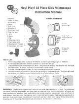
INTRODUCTION
TO
A
MICROSCOPIC
WORLD
In
this world there
is
an
abundance
of
living things.
Some
are
large enough to
be
seen
with
our own
eyes,
but
a lot
of
others
are
so
minute
that
millions
can
be
squeezed on the head
of
a pin.
Of
course these
tiny
organisms
can
only be
seen
through a microscope.
The microscope
was
invented many years ago. Since
then
it
has
opened a wider field
of
research, involving
many things
as
fascinating and
as
beautiful
as
you
can
imagine. Today, from
the
most elementary study
of
biology
to
the highly specialized fields
of
physiology,
some form
of
microscope
has
to
be used for
the
students
to
understand
better
the elaborate,
complicated forms
of
either living organisms or static
materials, which make up our living environment.
Your microscope will be either a source
of
fun for a
long-time
hobby
or
an
opening
door
to
advanced
knowledge
in
various fields
of
science.
We
hope you
will enjoy using it.
1 Eyepiece
2 Film Holder
3
Viewer
Screen
4 Viewer Head
5 Reflecting Mirror
6
Shutter Lever
7
Film winder
8 Body Tube
9 Focusing Knob
(HANDLE)
1 0 Objective
Lens
11
Revolving Turret
12
Arm
13
Clip
14 Stage
15
Color Filter
16
Illuminator Lamp
17
Mirror
18
Base
(BATTERY
CASE)
19
Condenser
Lens
Cap
2
INSTRUCTIONS
FOR
USE
OF
MICROSCOPE
Insert Zoom eyepiece into
the
bodytube
of
microscope and
tighten
the screw at the back
of
the tube.
If
the viewer
head
is
already in place,
remove
it
by loosening
the screw (refer
to
"USE
OF
THE
VIEWER")
2
Tilt
the
arm
of
the
microscope and adjust
the position
of
the mirror
so
that
the
light
is
reflected
through
the
stage hole. When
the
light
can
be
seen
through
the
eyepiece,
the
microscope
is
ready for
observation.
3 Put the prepared slide on
the stage and fix
it
in
place
with
the clips.
4
Now
decide which
magnification
to
use.
The
longer the length
of
the
objective
lens,
the greater
is
the magnification.
Usually observation
begins
with
a low setting.
5
If
you
want
to
change
magnification,
turn
the
revolving turret until you
feel a click.
6 Turn the focusing knob
to
lower
the lens before
touching the slide. Look
through the eyepiece.
Turn the knob in the other
direction until the image
comes into focus.
7
If
the
light
is
insufficient,
or
if
the sight
is
not
clear
at a high magnification, turn the illuminator lamp. Light
will
be
emitted automatically
to
enable observation.
8 The
light
source lamp
is
enabled
with
2 'AA'(LR6)
batteries being
put
inside the
base
as
illustrated.

ZOOM
MODELS:
The figure 1
OX.-..
20X
is
indicated on the
top
of
the Zoom
eyepiece.
By
rotating the silver knurled ring in clockwise direction, the
eyepiece power will zoom
to
20X.
Conversely,
if
you rotate
counterclockwise, the power will zoom
to
1
OX.
Assuming
that
you
are
using
an
objective turret
of
1
OX.
With
the eyepiece at 1
OX,
the magnification
power
of
the
combination becomes 1
Ox1
0=1
00.
The object you
see
is
enlarged 100 times.
If
you rotate the zoom eyepiece
to
20X, you
now
have a
combination
of
1 Ox20=200.
ZOOM
EYEPIECE
10x-20x
USE
OF
THE
VIEWER
Loosen the screw under
the
eyepiece in
counterclockwise
direction. Replace the
eyepiece
with
the
viewer and
tighten
the
screw.
To
increase the
brightness
of
the image,
cover the
Illuminator
Lamp
with
Condenser
Lens
Cap (provided in
the
set).
NEVER
USE
THE
CONDENSER
LENS
CAP
WHEN
YOU
ARE
LOOKING
THROUGH
THE
EYEPIECE.
Insert the plastic
viewer screen into the slot on
top
and lift the reflective
mirror
(as
shown in the picture above).
2 Carefully
put
prepared slide on the stage and beneath
the clips. Adjust the positions
of
the slide and the
illuminator such that the largest amount
of
light goes
through the stage hole onto the objective lens. While
moving the body tube by the focusing knob, look at the
viewer screen
to
check for a clear projection
of
the
sample. If the image on the screen
is
too
dark, adjust the
light source lamp, which should
not
be turned over 180
•.
3 Observation
of
the sample can be made
without
interruption for about
an
hour, due
to
the life
time
of
battery. When the microscope
is
not
used,
turn
off
the
illuminator lamp by flipping the mirror side upward.
3
HOW
TO
USE
AS
A
PROJECTION
DEVICE
Take
the viewer screen
out
of
the viewer head and face
it
toward
white
paper
or
a
white
wall then you can
see
projected image. When you project the image, please
do
it
in
the
dark room.
You
can
see
a clear image projected
approximately one meter distance.
HOW
TO
USE
THE
DRAWING
DEVICE
Adjust the arm
to
upright
position. After the image
has
been projected on the screen, eliminate the light
in
the
room.
2 Pull
out
the plastic viewer screen, and place a piece
of
white
paper horizontally in
front
of
the base
of
the
microscope. Position the reflecting mirror
as
shown in
the picture. Adjust the focusing knob until a satisfactory
image
is
projected.
Trace
the projected image with the
help
of
a pencil.
The
size
.
of
projected image
is
about
70
mm dia.
)
)

HOW
TO
TAKE
THE
MICROPHOTO
Follow
through
the
procedures
of
"USE
OF
THE
VIEWER"
on
previous page.
2
Without
the
condenser lens cap
on the
light
source
lamp,
turn
the
focusing knob slowly
until a clear image
is
projected on
the
viewer screen.
3
In
the direction
of
the
arrows in the picture
insert the 110 film
pack
into
the
film
holder
as
shown.
Without the condenser lens cap on the light source lamp,
turn
the focusing knob slowly
until
a clear image
is
projected on the viewer screen.
In
the direction
of
the arrow mark on the film holder, turn
the film winder
to
wind up the film.
You
will
see
the
arrow mark and film
number
by means
of
a small
rectangular
window
on the 110 film pack
Set
the inner 2 figures
out
of
the 4 figures in the
rectangular window.
4 Hold the microscope
base
with
left hand and
lift
the
shutter lever and release. Good results
can
be obtained
only
with
the correct exposure time.
Since
the required
time
depends on the
amount
of
light, color,
magnification, density
of
specimen and film speed, there
is
no specific time. However, testing on exposure
time
was
done in our lab, using slides
of
varying densities; and
the best results lies within range
of
0.5-1.5
sec.
5 Write the item's name, magnification and other reference
on the prepared slides. These information will become
useful when you take photographs next time.
HOW
TO
MAKE A
PREPARED
SLIDE
If the given sample
is
not
thin and transparent,
it
cannot be
observed by the microscope
as
the light from the reflector
or
the light source does
not
pass
though it. Fibres, pollen, wool,
or salt
can
be observed easily and cover
glass
is
optional.
Clear samples
are
stained first
with
a drop or
two
drops
of
methylene blue,
Eosin
or
other dyeing solutions available on
the market. (Note: These
are
dyeing solutions and therefore
could
cause
staining
of
clothing, carpets, and fabrics. Special
care should be taken when handling these solutions.
Also please read page 1 again
"EXPLANATION
OF
BOTTLES",
about
the
safety
information
associated
with
these
solutions.)
4
1) Temporary mount
Wipe the slide and cover glass clean. Thin sample
with
a
razor blade or the micro-slicer (Note: This process should be
under
an
adult's supervision. The blade
is
very sharp and
has
to
be handled
with
extreme caution. Please refer
to
following section for
use
of
micro-slicer).
Pick
up thinned
sample
with
tweezers and
put
it
on the centre part
of
the
glass slide. Put one drop
of
water on the sample
with
a
dissecting needle,or
if
the sample
is
clear,
use
one drop
of
the above mentioned dyeing solutions (Note:
The
needle
has
a sharp
point
so
handle
with
extreme caution) and then
gently
put
on cover
glass.
Avoid to trap any air bubbles.
Remove any
excess
water
or
dyeing solution
with
blotting
paper. Now
it
is
ready for observation. (Remember
to
wash
your hands after doing the preparation and remember
to
dispose the dyeing solutions according
to
the instructions
given on page 1
"ADVICE
FOR
SUPERVISING
ADULTS")
2) Permanent mount
Wipe the slide and cover
glass
clean
as
above (Temporary
Mount). Proceed
as
above. However before covering the
slide
with
the cover
glass,
add a few drops
of
gum media (not
included in the set- available from store) or Canada balsam
solution or transparent adhesive glue
with
a dissecting
needle
to
the
slide.
Push
down
on the cover glass
with
tweezers to fix
it
in place and leave
to
dry for about a
day.
HOW
TO
USE
THE
MICRO-SLICER
Put the specimen which you want
to
cut for study into the
holes
of
the micro-slicer (Note: the micro-slicer contains a
sharp edge
so
handle
with
extreme caution). Revolve the
blade. Then you can get
thin
slices
of
the specimen.

RAISING
A
FAMILY
OF
BRINE
SHRIMPS
(NOT
FOUND
IN
ALL
SETS)
It
is
relatively
easy
to
rear a batch
of
brine shrimps in the
home
laboratory. The
tiny
crustaceans are excellent
specimens for the microscope studies, being similar in many
ways
to
their bigger relatives, the lobsters, crabs, and crayfish.
Furthermore, the brine shrimps enrich the diet
of
aquarium
dwellers.
What makes the brine shrimp
easy
to
raise
is
that
its eggs
hatch in a relatively short time, from
twenty-four
to
forty-eight hours. The eggs
are
sold dried in small bottles or
vials. Dried eggs remain alive for five years or more
if
they
are
stored in a cool dry place. Almost every aquarium shop
carries a supply
of
the dried eggs
of
the brine shrimp.
In
order
to
hatch eggs, first
of
all float them in a container
of
sea
water. If
sea
water
is
unavailable, prepare a brine
solution by adding
two
teaspoons
of
table salt
to
a quart
of
water.
Sprinkle some brine shrimp eggs over salt solution and
allow
it
to
stand at room temperature
(70
-
80
F I 21-26
~
)
for a day
er two. The eggs will hatch into the nauplius larvae
which
is
the early life stage
of
most crustaceans.
To
bring the
larvae
to
maturity, transfer a small
number
to
another
container, for
if
they
are
left
to
fend for themselves in the
original culture they will begin
to
suffer from lack
of
oxygen
and die
off
shortly.
Prepare some fresh brine solution and add a small quantity
of
yeast to serve
as
food for the developing larvae. With a
pipette, transfer some
of
the new culture and allow
them
to
grow
to
maturity. Examination
of
the culture at frequent
intervals will reveal the entire life cycle
of
the brine shrimp,
Artemia salina.
Be
sure
to
observe the dried eggs, hatching
eggs, nauplius larvae, and the mature shrimp. When brine
shrimps are fed
to
aquarium dwellers, they should be
strained
out
of
the brine solution
with
a piece
of
fine
meshed cloth.
It
is
wise
to
wash the brine shrimp
with
fresh
water before
introducing
them
to
the aquarium
as
the
sudden increase in salt content may be harmful
to
fish.
5
OBSERVATION
OF
POLLEN
.
With a pair
of
tweezers, take
out
a piece
of
filament from the
flower. Slightly shake the filament
such
that a little amount
of
pollen fall
onto
the slide (too much pollen will make
observation difficult). At last, you may want
to
put
on the
glass
cover.
OBSERVATION
OF
FIBROUS
TISSUE
Try
to
get a piece
of
worn fabric or a piece
of
thread.
If
the
piece
of
fabric
is
torn or run, you may
see
some threads on its
edge.
Take
a closer look.
You
will discover
that
each piece
of
thread
can
be separated into small pieces, which
are
called
"filaments" or "fibres".
Place
a small quantity
of
fibres on a
blank slide and drip one drop
of
water on it
with
the point
of
a pipette.
You
can
put
on the cover
glass
and observe under
the microscope.

OBSERVATION
OF
SALTS
Fill a cylinder
with
warm water
until
the
water level
reaches 1/4
of
its
height
. Dissolve salt
into
the water. Add
more salt until the salt does
not
dissolve anymore. Swirl
the
cylinder constantly while dissolving
the
salt.
ci
::::-
1
2 With a small stick
or
a pipette, take
out
a small
amount
of
salt water and
drip
it
on a blank slide. Do
not
over drip.
3 Observe
how
crystals are gradually formed while viewing
through the microscope.
6
OBSERVATION
OF
STARCH
Ask
your parents
to
cut a
thoroughly
washed potato
into
2 pieces.
2
Rub
the
cut surface
of
the potato
with
a blank slide
as
it
is
illustrated below, until
white
liquid
is
formed on the
surface
of
the
glass.
3 With a stick or a pipette, take a small
amount
of
this
white
liquid from the slide and paste a very little
amount
on
the
center
of
the
blank slide. Make sure
the
stick
is
absolutely clean.
Cover
the
sample
with
a cover glass before observation
under the microscope.
/