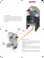
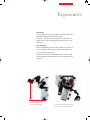
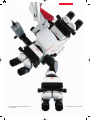
Leica DI C500 provides surgeons with outstanding color imaging and information technology capabilities during surgical procedures. It offers dual imaging with the ability to overlay data onto the surgical field, including CT/MRI scans, ultrasound, and navigation data. The device features a high display resolution, optimized beam paths, and flexible observation of data. It is designed for easy integration with existing surgical microscopes and supports a wide range of data applications.
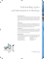
Leica DI C500 provides surgeons with outstanding color imaging and information technology capabilities during surgical procedures. It offers dual imaging with the ability to overlay data onto the surgical field, including CT/MRI scans, ultrasound, and navigation data. The device features a high display resolution, optimized beam paths, and flexible observation of data. It is designed for easy integration with existing surgical microscopes and supports a wide range of data applications.


-
 1
1
-
 2
2
-
 3
3
-
 4
4
-
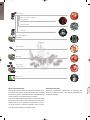 5
5
-
 6
6
-
 7
7
-
 8
8
-
 9
9
-
 10
10
-
 11
11
-
 12
12


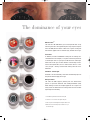
Leica DI C500 provides surgeons with outstanding color imaging and information technology capabilities during surgical procedures. It offers dual imaging with the ability to overlay data onto the surgical field, including CT/MRI scans, ultrasound, and navigation data. The device features a high display resolution, optimized beam paths, and flexible observation of data. It is designed for easy integration with existing surgical microscopes and supports a wide range of data applications.
Ask a question and I''ll find the answer in the document
Finding information in a document is now easier with AI
Related papers
-
Leica M525 OH4 Owner's manual
-
-
-
-
-
Leica Microsystems M165 C User manual
-
-
-
 Z Microsystems MACROFLUO User manual
-

Other documents
-
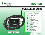 intwine connect ICG-150 User guide
-
 Steris Uroseal Adjustable Endoscopic Valve Operating instructions
-
 Intuitive Surgical 2AAZF-CHB01 User manual
-
 INDOORCYCLING Pairing of ANT+ Console & RPM transmitter User manual
-
Leica Microsystems M205 FCA User manual
-
-
Leica Microsystems LED2500 Application Note
-
-
Leica Microsystems FluoCombi III Application Note
-


