Page is loading ...

Art.No. 90-39100
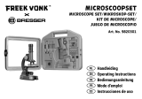
MIKROSKOP
MICROSCOPE
40X-1024X
Bedienungsanleitung
Operating instructions
Mode d’emploi
Handleiding
Istruzioni per l’uso
Instrucciones de uso
Руководство по эксплуатации
DE
EN
FR
NL
IT
ES
RU

12
General Information
About This instruction Manual
Please read the safety instructions in this manual carefully. To avoid damage to the unit
and the risk of injury, only use this product as described in the manual. Keep the instruc-
tion manual handy so that you can easily look up information on all the functions.
CAREFUL!
You will find this symbol before every section of text that deals with
the risk of minor to severe injuries resulting from improper use.
Intended Use
This product is intended only for private use.
It was developed for magnified viewing of natural and man-made objects.
General Warning
DANGER!
Tools with sharp edges and points are often used when working with this de-
vice. Because there is a risk of injury from such tools, store this device and all
tools and accessories in a location that is out of the reach of children.
DANGER!
This device contains electronic components which operate via a power source
(power supply and/or batteries). Only use the device as described in the man-
ual, otherwise you run the risk of an electric shock.
DANGER!
Do not expose the device to high temperatures. Use only the supplied power
supply or the recommended batteries. Do not short-circuit the device or batter-
ies or throw them into a fire! Excessive heat or improper handling could trigger
a short-circuit, a fire or an explosion. Never bend, pinch or pull the power and
connecting cables, extensions and adapters. Protect the cables from sharp
edges and heat. Before operating, check the device, cables and connections
for damage. Never use a damaged unit or a unit with damaged power cables.
Damaged parts must be exchanged immediately by an authorised service cen-
tre.
DANGER!
Children should only use the device under adult supervision. Keep packaging
material, like plastic bags and rubber bands, out of the reach of children, as
they pose a risk of choking.
CAREFUL!
Children must not have access to the included chemicals and liquids. Do not
drink the chemicals. Wash hands thoroughly with running water after using the
chemicals. In the event that the chemicals come into contact with your eyes or
mouth, rinse thoroughly with water. If you are in pain after exposure, contact a
doctor immediately and show him the substances.
NOTE!
Do not disassemble the device. In the event of a defect, please contact your
dealer. The dealer will contact the Service Centre and can send the device in to
be repaired, if necessary.
Do not expose the device to temperatures above 45 °C.

13
EN
Operating instructions
Parts overview:
B 10X WF eyepiece
C 16X WF eyepiece
D Barlow lens
E Electronic eyepiece (MicrOcular)
F Microscope head
G Objective revolver
H Microscope stage
I Focus wheel
J LED lighting (transmitted light)
1) Electricity supply
1! Microscope base
1@ Main plug
1# 5 slides, 10 covering glasses and 5 preparations in a plastic box
1$ Colour filter disc
1% LED lighting (reflected light)
1^ Direct light/transmitted light switch
1& Microscope tools: a) pipette; b) tweezers
1* Hatchery
1( MicroCut
2) Specimens: a) yeast; b) gum media (specimen inclusion medium); c)
sea salt; d) brine shrimp eggs
2! Locking screw
2@ Photomizer Software
1� General/Location
Make sure you position your microscope on a stable, solid surface. An electricity
supply is required for observation with the electric illuminator.
Position your device so that it can be disconnected from the power supply at any
time. The wall you use socket should be located near the device and easily ac-
cessible, since the plug on the power cord serves as a disconnecting device for
the power supply. Always pull on the plug to separate the device from the power
supply. Never pull on the cord.
2� Electric LED lighting
Before use, make sure the light switch (16) is set to 'off'.
The microscope has two light sources. Lighting can be of three types. Set the switch
(16) to 'II' to light the specimen from above (reflected light) or 'I' to light it from below
(transmitted light). Use setting 'III' to light the specimen simultaneously by transmit-
ted and reflected light. The transmitted light unit (9) is used for transparent speci-
mens (those on glass slides). To view solid, non-transparent specimens, use the
reflected light unit (15). Use of both forms of lighting simultaneously is only recom-
mended for semitransparent specimens. This operating mode is not recommended
for transmitted light specimens on slides as it may cause reflection on the slide.
3� Colour filter disc
The colour filter (14) under the microscope table (7) aids in viewing very bright and
transparent objects. Just select the right colour for the specimen in question. The
components of colourless or transparent objects (e.g. starch particles, single-cell
specimens) can thus be better recognised.
4� Microscope setup
The microscope head (5) will now be prepared for your first observation. First, loosen
the screw (21) and rotate the head into a convenient position. Begin every observation
with the lowest magnification. Place the microscope’s table (7) with the focus knob (8)
into the lowest position and rotate the objective revolver (6) until it locks on the lowest
magnification (4X).

14
NOTE:
Make sure to place the microscope's table (7) in its lowest
position before changing the objective in order to prevent
damage to the microscope.
Insert the 10X eyepiece (No. 1, 1) in the Barlow lens (No. 1, 3).
Take care that the Barlow lens is inserted completely into the monocular head (No.
1, 4).
5� Observation
After you have set up the microscope with the proper illumination, the following
principles are important:
Begin each observation at the lowest magnification, so that the centre and position
of the object to be viewed is in focus. The higher the magnification, the more light
is required for good picture quality.
Place a permanent slide culture directly under the microscope lens on the plate (7).
The specimen to be examined must be directly over the lighting.
Look through the eyepiece (1 and 2) and carefully turn the focus wheel (8) until you
can see a sharp picture.
Now you can progress to a higher magnification. Slowly pull the Barlow lens (No.
2, 3) out of the monocular barrel (No. 2, 4). When the Barlow lens is nearly entirely
pulled out, the magnification is raised to 2X.
For even higher magnification, you can put the 16X eyepiece (2) into the objec-
tive revolver (6) and rotate the objective revolver to a higher magnification (10X or
40X).
i
TIP:
Depending on the preparation, higher magnifications do not
always lead to better pictures.
When changing the magnification of your microscope by changing or adjusting the
eyepiece, objective lens or Barlow lens, you must readjust the focus wheel (8) to
sharpen the image.
NOTE:
Please be very careful when doing this. If you move the mechani-
cal plate upward too fast, the objective lens and the slide can
touch and become damaged.
6� Condition and prepare viewed objects
6�1� Condition
With the Barlow lens nearly fully extended, your microscope's magnification can
be doubled. Both transparent and non-transparent specimens can be examined
with this microscope, which features both direct and transmitted light. If opaque
specimens are being examined, such as small animals, plant parts, tissues, stones
and the like, the light is reflected from the specimen, through the lens and eye-
piece, where it is magnified, to the eye (reflected light principle, switch position
I). If opaque specimens are being examined, the light from below goes through
the specimen, lens and eyepiece to the eye and is magnified en route (direct light
principle, switch position II).
Some small water organisms, plant parts and animal components are transparent
by nature, but many others require pretreatment — that is, you need to make a thin-
nest possible slice of the object by hand cutting or using a microtome, and then
examine this sample.
6�2� Creation of thin preparation cuts
Specimens should be sliced as thin as possible. A little wax or paraffin is needed to
achieve the best results. Put the wax into a heat-safe bowl and heat it over a flame
until the wax is melted. You can use a candle flame to melt the wax.

15
EN
DANGER!
Be exremely carfeful when dealing with hot wax, as there is a danger
of being burned.
Then, dip the specimen several times in the liquid wax. Allow the wax that encases
the specimen to harden. Use a MicroCut (19) or other small knife or scalpel to
make very thin slices of the object in its wax casing.
DANGER!
Be extremely careful when using the MicroCut, knife or scalpel.
These instruments are very sharp and pose a risk of injury.
Place the slices on a glass slide and cover them with another slide before attempt-
ing to view them with the microscope.
6�3� Creation of your own preparation
Put the object to be observed on a glass slide and cover the object with a drop of
distilled water (No. 3) using the pipette (No. 3, 17a).
Set a cover glass (available at a well-stocked hobby shop) perpendicular to the
edge of the water drop, so that the water runs along the edge of the cover glass
(No. 4). Now lower now the cover glass slowly over the water drop.
i
TIP:
The gum medium (20b) supplied is used to make permanent slide
cultures. Add it instead of distilled water.
The gum medium hardens so that the specimen is permanently
affixed to its slide.
7� Experiments
Now that you're familiar with your microscope's functions and how to prepare
slides, you can complete the following experiments and observe the results under
your microscope.
7�1� Newspaper print
Objects:
1. A small piece of paper from a newspaper with parts of a picture and some
letters
2. A similar piece of paper from an illustrated magazine:
Use your microscope at the lowest magnification and make a slide preparation
from each object. Place the slide with the newspaper on the microscope table and
observe the slide. The letters in the newspaper appear broken because the news-
paper is printed on raw, inferior paper. Now observe the slide with the magazine
preparation. Letters of the magazine appear smoother and more complete. The
picture from the newspaper consists of many small points, which appear somewhat
dirty. The pixels (raster points) of the magazine image appear sharper.
7�2� Textile fibres
Objects and accessories:
1. Threads of different textiles: Cotton, linen, wool, silk, Celanese, nylon and
any others you can find.
2. Two needles:
Put each thread on a glass slide and fray each with the help of the two needles. Put
a drop of water over each thread with the pipette and cover each with a cover glass.
Adjust the microscope to a low magnification. Cotton fibres are of plant origin and
look, under the microscope, like a flat, twisted band. The fibres are thicker and
rounder at the edges than in the centre. Cotton fibres consist primarily of long, col-
lapsed tubes. Linen fibres are also of plant origin; they are round and run in straight
lines. The fibres shine like silk and exhibit numerous swellings along the shaft of
the fibre. Silk is of animal origin and consists of solid fibres of smaller diameter than
the hollow vegetable fibres. Each silk fibre is smooth and even and has the appear-
ance of a small glass rod. Wool fibres are also of animal origin; the surface consists
of overlapping scales, which appear broken and wavy. If possible, compare wool
fibres from different weaving mills, and note the differences in the appearance of
the fibres. Experts can determine the country of origin of wool based on its appear-
ance under a microscope. Celanese is artificially manufactured by a long chemical
process. All Celanese fibres show hard, dark lines on a smooth, shining surface.

16
The fibres crinkle in the same way after drying. Observe the similarities and differ-
ences between the different fibres.
7�3� Saltwater brine shrimps
Accessories:
1. Brine shrimp eggs (20d)
2. Sea salt (20c)
3. Hatchery (18)
4. Yeast (20a)
CAREFUL!
These eggs are not fit for human consumption.
7�3�1� Winter eggs of Artemia salina
Artemia salina are species of brine shrimp typically found in salt lakes — bodies of
water with a higher salinity than even the ocean. During a drought, a salt lake can
become a hostile habitat for organisms, and entire populations of Artemia salina
sometimes die off. During drought conditions, to ensure that the species will re-
populate the salt lake when the drought ends, Artemia salina lay thick-shelled eggs
called winter eggs that can survive for up to ten years in a dormant state. Winter
eggs can withstand heat, cold and chemicals. These eggs hatch when favourable
conditions return to their ambient environment. The eggs provided (20d) are of
this type.
7�3�2� Hatching winter eggs
To hatch the brine shrimps, create a solution with an appropriate salinity and tem-
perature. First, fill two containers with a half litre of freshwater each, and let them
both stand for about thirty hours. Next, pour half of the provided salt (20c) into one
container and stir the solution until the salt dissolves. Pour some of this solution
into the hatchery (18). Place a few eggs close to the lid. Position the hatchery
somewhere with plenty of light but not in direct sunlight. The ambient temperature
should ideally hover around 25 °C. As water in the hatchery evaporates, gradually
add freshwater from the second container. After two to three days, the eggs will
hatch brine shrimp larvae, called nauplii.
7�3�3� Observing Artemia salina under a microscope
Using the pipette (17a), move some larvae from the container to a microscope slide
for examination. When viewing the larvae, you’ll notice that they swim through the
solution using hairlike limbs! Each day, examine a few more. You can even view the
entire hatchery under the microscope if you remove its lid. The larvae will mature in
six to ten weeks, depending on the temperature of the water. Soon, you will have
an entire generation of saltwater brine shrimps that reproduce frequently!
7�3�4� Feeding your Artemia salina
Feed your brine shrimps often to keep them alive. The best food is dry powdered
yeast (20a). Give them some every other day. Be careful not to overfeed them, as
doing so can cause the water to stagnate and poison the brine shrimps. If the water
does begin to stagnate (you’ll see it darkening), transfer the brine shrimps to the
fresh saline solution you have prepared earlier (see 7.3.2).
8� MicrOcular setup
NOTE:
The MicrOcular only works without the Barlow lens supplied.
Magnification setting is changed by using the MicrOcular and must
be reset by re-focussing.
Remove the Barlow lens (Fig 5, 3) and eyepiece currently in use from the eyepiece
holder (Fig 5, 5) and replace them with the MicrOcular (PC-Ocular) (Fig 6, 4) and
reducer lens (Fig 6, F) as in illustration 6 in the holders (Fig 6, 5).

17
EN
NOTE:
Please do not yet connect MicrOcular and PC. Please go through
the items below in sequence.
9� Installation and use of the software
9�1� Software and installation information
A software CD is included with your microscope. The software and drivers on this
CD must be installed on your computer in order to use your MicrOcular. Once you
connect the MicOcular to your computer after installation, you can view pictures on
and save them to your computer using Photomizer. To install the software and driver
correctly, simply follow the installation steps.
9�2� Software installation
1. Important: Before inserting the CD, first plug the USB cable into the USB port
on your PC. Windows will now recognize the new device, and will indicate this in
a notification window. Now please click on “Cancel”.
2. Now insert the CD-ROM that came in your package into the CD/DVD drive of
your computer. The installation menu starts automatically. If it does not, go to the
Windows explorer and select the CD/DVD drive (most of the time, it’s the “D”
drive, but it can have another letter). From there, start the file “autorun.exe” by
double-clicking with the left mouse button.
9�2�1� Driver installation
To install the driver software, click on the menu point “Install Driver” with the
mouse cursor. Then follow the installation program instructions. During the soft-
ware installation the correct driver for your operating system will be installed au-
tomatically. No manual input is needed. In rare cases the device may not be rec-
ognised by your computer. As a rule you need then only install the driver manually
from the CD. If this fails please refer to the troubleshooting chapter that follows.
9�2�2� Installation of the Image Editing Software Photomizer
The image editing software “Photomizer” is located on the software CD. You can
edit your pictures here.
1. To install the software, click once on the menu point “Install PHOTOMIZER”
with the left mouse button.
2. The Photomizer Software requires Microsoft .NET Framework 4.0, which it will
install if it is not already on your system. If it is already on your system, you may
skip to step 5.
3. In the welcome window, please accept the Microsoft license agreement, and
then click “Install”. The installation can take a few minutes.
4. As soon as everything is installed, click “Finish”.
5. Now you will be presented with a choice, in which you can choose your
language.
Make your selection and confirm it by clicking „OK“.
6. When you see “Welcome“, click on „Next“.
7. In the next window, you will be asked for the „Destination Folder“. Here, just
click on „Next“.
8. Now, the window with the Setup-Status will appear – here, a progress bar will
inform you about the ongoing installation. This process can take a few minutes.
9. The window “Photomizer is being installed” appears. Click on “Finish”. The
installation ends.

18
i
TIP:
To use the MicrOcular long-term we recommend to
always connect it to the same USB port.
10� Using the MicrOcular
10�1� Preparation
1. Slide a specimen under your microscope and focus on it.
2. Remove the eyepiece and Barlow lens from the eyepiece support and the dust
cap from your MicrOcular and install same in the eyepiece supports instead of
the Barlow lens.
3. Start your PC if you haven‘t yet and connect your MicrOcular to the USB port of
your computer.
10�2� Showing and saving MicrOcular images on your PC
1. Start the Photomizer Software.
2. Click „Open camera“
3. In the event that you have connected more than one unit, you can choose the
desired unit in the subsequent selection. Click on "USB 2.0 Webcam". If only
one device is connected, this step is omitted.
4.
The camera image should now be visible on your screen. Focus the microscope
image.
5. Click „Capture“ to record an image. It will then be shown on the right.
6. Click on it to select it and then click „Transfer image“.
7.
This takes you to the Photomizer software.
8. File - Save as
10�3� The Photomizer Software
If you need help in the „Photomizer“ programme click „?“ and then „Open help“. If
you need further assistance please visit the maker‘s home page at www.photomizer.
net/bresser
Care and maintenance
Before cleaning, separate the device from the power supply by removing the plug.
Only use a dry cloth to clean the exterior of the device.
NOTE:
Do not use any cleaning fluid to avoid damaging the electronics.
Clean the lenses eyepieces and lenses only with a soft, lint-free cloth, like a mi-
crofibre cloth.
NOTE:
Do not apply excess pressure to the cloth so as to avoid scrat-
ching the lenses.
To remove more stubborn dirt, moisten the cleaning cloth with an eyeglass-clean-
ing solution and wipe the lenses gently. Protect the device from dust and moisture.
After use, particularly in high humidity, let the device acclimatize for a short period
of time, so that the residual moisture can dissipate before storing.
Troubleshooting
Problem Solution
No picture visible • switch light on
• adjust focus
Picture flickers • if necessary,
while viewing
adjust resolution of the
with PC eyepiece video graphics board

19
EN
Software installation • confirm by
reports clicking “OK”
“not XP approved”
System requirements for PC eyepiece
Minimum system requirements: PC with an Intel Pentium IV processor or higher;
Windows XP with Service Pack 3*, Windows Vista (32/64Bit) with Service Pack 2*
or Windows 7 (32/64Bit) with Service Pack 1*; .NET Framework 4.0*; min. 1024
MB RAM (64Bit = 2048 MB); min. 500 MB free hard drive space; free USB port;
CD/DVD/BD drive.
* available for free via Windows Update (Internet connection needed)
Magnification table
Eyepiece Objective Magnification with Barlow lens
10X 4X 40X 64X
10X 10X 100X 160X
10X 40X 400X 640X
16X 4X 64X 102X
16X 10X 160X 256X
16X 40X 640X 1024X
DISPOSAL
Dispose of the packaging materials properly, according to their type, such as
paper or cardboard. Contact your local waste-disposal service or environmen-
tal authority for information on the proper disposal.
Do not dispose of electronic devices in the household garbage!
As per Directive 2002/96/EC of the European Parliament on waste elec-
trical and electronic equipment and its adaptation into German law, used
electronic devices must be collected separately and recycled in an environmentally
friendly manner.
In accordance with the regulations concerning batteries and rechargeable
batteries, disposing of them in the normal household waste is explicitly forbid-
den. Please make sure to dispose of your used batteries as required by law — at a
local collection point or in the retail market. Disposal in domestic waste violates the
Battery Directive.
Cd¹ Hg² Pb³
1
battery contains cadmium
2
battery contains mercury
3
battery contains lead
Declaration of Conformity
Product Type: Microscope
Product Name: Microscope 40X-1024X
Article No.: 90-39100
Bresser GmbH has issued a 'Declaration of Conformity' in accordance with appli-
cable guidelines and corresponding standards. This can be viewed any time upon
request.

ANL9039100MSP0714NG
MIKROSKOP
MICROSCOPE
40X-1280X
© 2014 National Geographic Society
NATIONAL GEOGRAPHIC and Yellow Border Design
are trademarks of the National Geographic Society,
used under license. All rights reserved.
Visit our website:
www.nationalgeographic.com
Irrtümer und technische Änderungen vorbehalten.
Errors and technical changes reserved.
Bresser GmbH
Gutenbergstr. 2 · DE-46414 Rhede
www.bresser.de · info@bresser.de
/