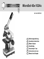
Bresser Junior Biolux CA 40x-1024x Microscope incl. Smartphone Holder Owner's manual
- Category
- Microscopes
- Type
- Owner's manual

MicroSet 40x-1024x
Art. No. 88-55002
DE
Bedienungsanleitung
GB
Operating Instructions
FR
Mode d’emploi
NL
Handleiding
IT
Istruzioni per l’uso
ES
Instrucciones de uso
PT
Manual de utilização

DE
Bedienungsanleitung ................................... 4
GB
Operating Instructions ............................... 10
FR
Mode d’emploi ............................................ 16
NL
Handleiding .................................................22
IT
Istruzioni per l’uso ......................................28
ES
Instrucciones de uso ..................................34
PT
Manual de utilização ..................................40
ACHTUNG!
Beinhaltet funktionale scharfkantige
Ecken und Punkte! Kleine Teile,
Erstickungsgefahr. Nicht für Kinder
unter 3 Jahren geeignet.
WARNING!
Contains functional sharp edges and
points. Choking hazard – small parts.
Not for children under three years.
0-3
Page is loading ...
Page is loading ...
Page is loading ...
Page is loading ...
Page is loading ...
Page is loading ...
Page is loading ...

10
RISK to your child.
Aids with sharp edges and tips are so-
metimes used with this device. Please
store the device and all of its accessories and
aids out of the reach of children. There is a
risk of
INJURY.
This device contains electronic components
that are powered by either a mains connec-
tion or batteries. Never leave a child unsu-
pervised with this device. The device should
only be used as per these instructions other-
wise there is a serious
RISK of ELECTRICAL
SHOCK.
Children should only use this device under su-
pervision. Keep packaging materials (plastic
bags, rubber bands, etc.) away from children.
There is a risk of
SUFFOCATION.
The chemicals and liquids provided should
be kept out of reach of children. Do not drink
the chemicals! Hands should be washed
thoroughly under running water after use. In
case of accidental contact with the eyes or
mouth rinse with water. Seek medical treat-
ment for ailments arising from contact with the
chemical substances and take the chemicals
with you to the doctor.
FIRE-/ DANGER OF EXPLOSION!
Do not expose the device to high tem-
peratures. Use only the mains adapter
supplied or those battery types recommen-
ded. Never short circuit the device or batte-
ries or throw into a fi re. Exposure to high tem-
peratures or misuse of the device can lead to
short circuits, fi re or even explosion!
RISK of material damage
Never take the device apart. Please
consult your dealer if there are any
defects. The dealer will contact our service
centre and send the device in for repair if nee-
ded.
Position your device so that it can be discon-
nected from the power supply at any time. The
wall socket should always be located near the
device and be easily accessible, since the
plug on the power cord serves as a discon-
necting device for the power supply.
Do not subject the device to temperatures ex-
ceeding 60° C.
TIPS on cleaning
Remove the device from it’s ener-
gy source before cleaning (remove
plug from socket / remove batte-
ries).
Clean the exterior of device with a dry cloth.
Do not use cleaning fl uids so as to avoid
causing damage to electronic components.
Clean the lens (objective and eyepiece) only
with the cloth supplied or some other soft lint-
free cloth (e.g. micro-fi bre). Do not use ex-
cessive pressure - this may scratch the lens.
Dampen the cleaning cloth with a spectacle
cleaning fl uid and use it on very dirty lenses.
Protect the device from dust and moisture.
Store the device in the bag supplied or in its
original packaging. Batteries should be re-
moved from the device if it is not going to be
used for a long period of time.

11
GB
DISPOSAL
Dispose of the packaging material/s as
legally required. Consult the local au-
thority on the matter if necessary.
Do not dispose of electrical equipment
in your ordinary refuse. The European
guideline 2002/96/EU on Electronic
and Electrical Equipment Waste and relevant
laws applying to it require such used equip-
ment to be separately collected and recycled
in an environment-friendly manner. Empty bat-
teries and accumulators must be disposed of
separately. Information on disposing of all
such equipment made after 01 June 2006
can be obtained from your local authority.
EC Declaration of Conformity
Bresser GmbH has issued a ‘Decla-
ration of Conformity’ in accordance
with applicable guidelines and corresponding
standards. This can be viewed any time upon
request.

12
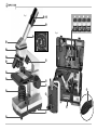
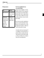
Here are the parts of your microscope
(Fig. 1-6):
1 10x WF Eyepiece
2 16x WF Eyepiece
3 Barlow Lens
4 Compartment for optional accessories
5 Eyepiece supports
6 Microscope Head
7 Set Screw
8 Objective Nosepiece
9 Objective
10 Clips
11 Microscope Stage
12 LED Illumination (transmitted light)
13 Microscope Base
14 Selection Knob for Illumination
15 Battery compartment (3x AA)
16 Focus knob
17 Color Filter
18 LED Illumination (refl ected light)
19 Compartment for optional accessories
20 5 Slides, 10 Cover Sips and 5 Prepared
Specimens plastic box
21 Specimens:
a) Yeast
b) “Gum Media” Glue
c) Sea Salt
d) Shrimp Eggs
22 Smartphone holder
23 Specimen Slicer
24 Shrimp Hatchery
25 Tweezers
26 Pipette
27 Carrying Case
How do I use my microscope?
Before you assemble your microscope, make
sure that the table, desk or whatever surface
that you want to place it on is stable, and does
not wobble.
How do I operate the electric LED
illumination?
The batteries supplied should be inserted into
the battery compartment (15) located in the
base plate before use.
Remove the battery compartment cover by
pressing lightly on the cover clip. Insert the
batteries (3x AA) into the holder.
There are two lights on the microscope. They
do not use light bulbs, but rather light-emitting
diodes (LED). The fi rst lamp shines onto the
specimen from below and the second from
above. (The thing that you want to observe
with the microscope is called the object or
specimen, by the way.) You can use each
lamp on its own, or both of them together.
There is a selection knob for this (Fig. 1, 14).
It has three numbers: I, II and III.
If you select the …
I, the light only comes from below (transmit-
ted light).
II, the light only comes from above (refl ected
light).
III, both lamps shine light on the specimen.
For transparent objects (transmitted-light ob-
jects), number I is best. In order to observe
fi rm, non-transparent objects (direct-light ob-
jects), select number II. For semi-transparent
objects, it is best to select number III.
It is not recommended to use number III for
transmitted-light objects on slides, since the
light may cause refl ections on the surface of
the slide, which will disturb your observation.
When do I use the color fi lters?
The color fi lters (Fig. 1, 17) are located below
the microscope stage (Fig. 1, 11). They help
you when you are observing very bright or
clear specimens. Here, you can choose from
various colors. This helps you better recog-
nize the components of colorless or transpar-
ent objects (e.g. grains of starch, protozoa).

13
GB
How do I adjust my microscope correctly?
First, loosen the screw (Fig. 1, 7) and turn the
microscope head (Fig. 1, 6) into a comfort-
able viewing position.
Each observation starts with the lowest mag-
nifi cation.
Adjust the microscope stage (Fig. 1, 11) so
that it goes all the way down to the lowest
position. Then, turn the objective nosepiece
(Fig. 1, 8) until it clicks into place at the lowest
magnifi cation (objective 4x).
Note:
Before you change the objective setting, al-
ways move the microscope stage (Fig. 1, 11)
to its lowest position. This way, you can avoid
causing any damage!
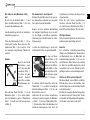
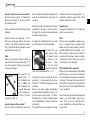
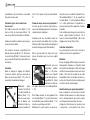
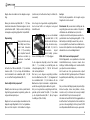
Now, insert the 10x
eyepiece (Fig. 1, 1)
into the Barlow lens
(Fig. 1, 3). Make sure
that the Barlow lens
is placed all the way
into the eyepiece sup-
ports (Fig. 1, 5) and is
not pulled out (Fig. 2).
How do I observe the specimen?
After you have assembled the microscope
with the adequate illumination and adjusted it
correctly, the following basic rules are to be
observed:
Start with a simple observation at the lowest
magnifi cation. This way, it is easier to position
the object in the middle (centering) and make
the image sharp (focusing).
The higher the magnifi cation, the more light
you will require for a good image quality.
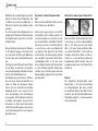
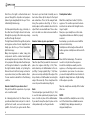
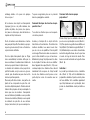
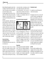
Now place the pre-
pared specimen (Fig.
6, 20) directly under
the objective on the
microscope stage
(Fig. 3). The object
should be located di-
rectly over the illumination (Fig. 1, 12).
In the next step, take a look through the eye-
piece (Fig. 1, 1) and carefully turn the focus
knob (Fig. 1, 16) until the image appears clear
and sharp.
Now you can select a higher magnifi cation
by slowly removing the Barlow lens (Fig. 1, 3)
from the eyepiece support (Fig. 1, 5). When
the Barlow lens is almost completely pulled
out, the magnifi cation can be increased to al-
most double.
If you would like an even higher level of mag-
nifi cation, insert the 16x eyepiece (Fig. 1, 2)
and turn the objective nosepiece (Fig. 1, 8) to
a higher setting (10x or 40x).
Important tip:
The highest magnifi cation is not always the
best for every specimen!
Note:
Each time the magnifi cation changes (eye-
piece or objective change, pulling out the
Barlow lens), the image sharpness must be
readjusted with the focus knob (Fig. 1, 16).
When doing this, make sure to be careful. If
you move the microscope stage too quickly,
the objective and the slide could come into
contact and become damaged!
Which light for which specimen?
With this unit, a refl ected light and transmitted
light microscope, you can observe transpar-
ent, semi-transparent as well as non-transpar-
ent objects. The image of the given object of
observation is “transported” through the light.
As a result, only the correct light will allow you
to see something!
If you are observing non-transparent (opaque)
objects (e.g. small animals, plant compo-
nents, stones, coins, etc.) with this micro-
scope, the light falls on the object that is be-
ing observed.
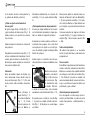
Fig. 3
I
2)
Fig. 2
D
f
B/C

14
From there, the light is refl ected back and
passes through the objective and eyepiece
(where it gets magnifi ed) into the eye. This is
refl ected light microscopy.
For transparent objections (e.g. protozoa), on
the other hand, the light shines from below,
through the opening in the microscope stage
and then through the object.
The light travels further through the objective
and eyepiece, where it is also magnifi ed, and
fi nally goes into the eye. This is transmitted-
light microscopy.
Many microorganisms in water, many plan
components and the smallest animal parts
are already transparent in nature. Others have
to be prepared. We may make them transpar-
ent through a treatment or penetration with
the right materials (media), or by taking the
thinnest slices from them (using our hand or
a specimen slicer), and then examine them.
You can read more about this in the following
sections.
How do I make thin specimen slices?
Only do this with the supervision of your par-
ents or another adult.
As I already pointed out, the thinnest slices
possible are taken from an object. In order to
get the best results, we need some wax or
paraffi n. It is best if you get a candle. Place
the wax in a pot and heat it carefully over a
low burner. Now, dip the object in the liquid
wax a few times. Then, let the wax get hard.
Using the specimen slicer (Fig. 6, 23) or a
knife/scalpel, cut the smallest slices from the
object that is covered with wax. These slices
are to be laid on a slide and covered with a
cover slip.
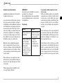
How do I make my own specimens?
Fig. 7 Fig. 8
2^
Take the object that you want to observe and
place it on a glass slide (Fig. 6, 20). Then,
add a few drops of distilled water on the ob-
ject (Fig. 7) using a pipette (Fig. 7, 26). Now,
place a cover slip vertically at the edge of the
drop of water, so that the water runs along the
edge of the cover slip. Then, slowly lower the
cover slip over the water drops (Fig. 8).
Note:
The included glue “gum media” (Fig. 5, 21b)
is used to make permanent prepared speci-
mens. Use this in place of the distilled water.
If you want to keep the object in place on the
slide permanently, use the gum media.
Smartphone holder
Attach the smartphone holder (22) to the
eyepiece. The suction cups must be clean
and free from dust and dirt. A slight moiste-
ning is helpful.
Now press your smartphone on the retai-
ning plate and make sure that it is properly
secured.
As a backup, you should secure it with the
enclosed rubber strap.
Smartphones with a rough surface will not
hold as good as smartphones with a smooth
surface.
Now start the Camera app. The camera
needs to rest just above the eyepiece.
Center the smartphone exactly over the
eyepiece, so the image can be seen precise-
ly centered on your screen. In some cases
you need to adjust with the zoom function to
display the image fullscreen. A light shading
at the edges is possible.
Take the smartphone carefully off the holder
after use.
NOTE:
Make sure that the smartphone can not slip
out of the holder. Bresser GmbH assumes
no liability for any damages caused by a
dropped smartphone.

15
GB
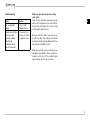
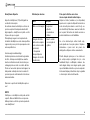
Troubleshooting
Error Solution
No recognizable
image
• Turn on light
• Readjust focus
Image fl ickers (while
observing with
optional available
MicrOcular)
= Monitor refresh
rate not adequate
• If necessary, in-
crease resolution
of graphics card
Make sure your microscope has a long
service life.
Clean the lens (objective and eyepiece) only
with the cloth supplied or some other soft lint-
free cloth (e.g.microfi bre). Do not press hard
as this might scratch the lens.
Ask your parents to help if your microscope
is really very dirty. The cleaning cloth should
be moistened with cleaning fl uid and the lens
wiped clean using little pressure.
Make sure your microscope is always protec-
ted against dust and dirt. After use leave it in
a warm room to dry off. Then install the dust
caps and keep it in the case provided.
Page is loading ...
Page is loading ...
Page is loading ...
Page is loading ...
Page is loading ...
Page is loading ...
Page is loading ...
Page is loading ...
Page is loading ...
Page is loading ...
Page is loading ...
Page is loading ...
Page is loading ...
Page is loading ...
Page is loading ...
Page is loading ...
Page is loading ...
Page is loading ...
Page is loading ...
Page is loading ...
Page is loading ...
Page is loading ...
Page is loading ...
Page is loading ...
Page is loading ...
Page is loading ...
Page is loading ...
Page is loading ...
Page is loading ...
Page is loading ...
Page is loading ...
-
 1
1
-
 2
2
-
 3
3
-
 4
4
-
 5
5
-
 6
6
-
 7
7
-
 8
8
-
 9
9
-
 10
10
-
 11
11
-
 12
12
-
 13
13
-
 14
14
-
 15
15
-
 16
16
-
 17
17
-
 18
18
-
 19
19
-
 20
20
-
 21
21
-
 22
22
-
 23
23
-
 24
24
-
 25
25
-
 26
26
-
 27
27
-
 28
28
-
 29
29
-
 30
30
-
 31
31
-
 32
32
-
 33
33
-
 34
34
-
 35
35
-
 36
36
-
 37
37
-
 38
38
-
 39
39
-
 40
40
-
 41
41
-
 42
42
-
 43
43
-
 44
44
-
 45
45
-
 46
46


Bresser Junior Biolux CA 40x-1024x Microscope incl. Smartphone Holder Owner's manual
- Category
- Microscopes
- Type
- Owner's manual
Ask a question and I''ll find the answer in the document
Finding information in a document is now easier with AI
in other languages
- italiano: Bresser Junior Biolux CA 40x-1024x Microscope incl. Smartphone Holder Manuale del proprietario
- français: Bresser Junior Biolux CA 40x-1024x Microscope incl. Smartphone Holder Le manuel du propriétaire
- español: Bresser Junior Biolux CA 40x-1024x Microscope incl. Smartphone Holder El manual del propietario
- Deutsch: Bresser Junior Biolux CA 40x-1024x Microscope incl. Smartphone Holder Bedienungsanleitung
- Nederlands: Bresser Junior Biolux CA 40x-1024x Microscope incl. Smartphone Holder de handleiding
- português: Bresser Junior Biolux CA 40x-1024x Microscope incl. Smartphone Holder Manual do proprietário
Related papers
-
Bresser Junior MicroSet 40x-1024x Owner's manual
-
Bresser Junior 8851200 Owner's manual
-
-
-
-
-
-
-
-
Other documents
-
National Geographic Biolux Student Microscope-Set Owner's manual
-
-
-
 Freek Vonk 9820301 Owner's manual
-
Bresser Biolux ICD 20x Stereo Microscope Owner's manual
-
-
-
-
Tasco Novice Telescope & Microscope 49TN / 5TN / 45T / 54TN User manual
-
